中国组织工程研究 ›› 2021, Vol. 25 ›› Issue (4): 537-541.doi: 10.3969/j.issn.2095-4344.2365

• 组织工程口腔材料 tissue-engineered oral materials • 上一篇    下一篇

E-Max瓷嵌体三维有限元模型粘接界面应力分析

张国梅1,祝  军1,胡  杨2,焦红卫3   

  1. 1上海市宝山区罗店医院口腔科,上海市   201908;2新疆医科大学第一附属医院口腔修复科,新疆维吾尔自治区乌鲁木齐市   830054;3上海市口腔病防治院,上海口腔医院,永嘉路特需门诊,上海市   200001
  • 收稿日期:2019-12-24 修回日期:2019-12-27 接受日期:2020-04-22 出版日期:2021-02-08 发布日期:2020-11-21
  • 通讯作者: 焦红卫,副主任医师,上海市口腔病防治院,上海口腔医院,永嘉路特需门诊,上海市 200001
  • 作者简介:张国梅,女,1978年生,黑龙江省鹤岗市人,汉族,硕士,主要从事口腔修复学研究。
  • 基金资助:
    新疆维吾尔自治区重点研发任务专项基金(2016B03049-2)

Stress of three-dimensional finite element models of E-MAX porcelain inlay

Zhang Guomei1, Zhu Jun1, Hu Yang2, Jiao Hongwei3   

  1. 1Department of Stomatology, Luodian Hospital of Baoshan District of Shanghai, Shanghai 201908, China; 2Department of Prosthodontics, First Affiliated Hospital of Xinjiang Medical University, Urumqi 830054, Xinjiang Uygur Autonomous Region, China; 3Cavity Prevention Center in Shanghai, Shanghai Stomatological Hospital, Yongjia Road Special Need Clinic, Shanghai 200001, China
  • Received:2019-12-24 Revised:2019-12-27 Accepted:2020-04-22 Online:2021-02-08 Published:2020-11-21
  • Contact: Jiao Hongwei, Associate chief physician, Cavity Prevention Center in Shanghai, Shanghai Stomatological Hospital, Yongjia Road Special Need Clinic, Shanghai 200001, China
  • About author:Zhang Guomei, Master, Department of Stomatology, Luodian Hospital of Baoshan District of Shanghai, Shanghai 201908, China
  • Supported by:
    the Special Fund for Key Research & Development Tasks of Xinjiang Uygur Autonomous Region, No. 2016B03049-2

摘要:

文题释义:
瓷嵌体:嵌体是一种临床应用悠久的口腔修复技术,是嵌入牙体内部用以恢复牙体缺损形态和功能的修复体,属于口腔固定修复体类,具有咀嚼效率高、磨牙量少、异物感较小等特点,金属类嵌体的机械强度高,E-Max瓷嵌体的美学性能和生物相容性较好,在前牙美学区和后牙应力负载较低部位有广阔的应用前景。
三维有限元模型:是指利用计算机数学算法对口颌系统(几何和载荷工况)进行模拟,利用简单而又相互作用的元素(即单元),用有限数量的未知量去推算无限未知量的真实系统,可模拟口颌系统咬合应力施加环境,便于分析咬合应力与修复体、粘接界面和剩余牙体组织的应力状况,优化E-Max瓷嵌体洞型设计和黏结剂选择。 

背景:E-Max瓷嵌体具有良好的美学、粘接与机械性能,在牙体缺损修复领域有广阔的应用前景。
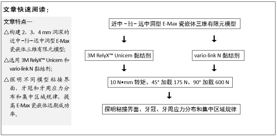
目的:构建不同黏结剂和不同洞深的近中-牙合-远中洞型E-Max瓷嵌体修复三维有限元模型,探究不同模型的应力分布和集中区域规律。
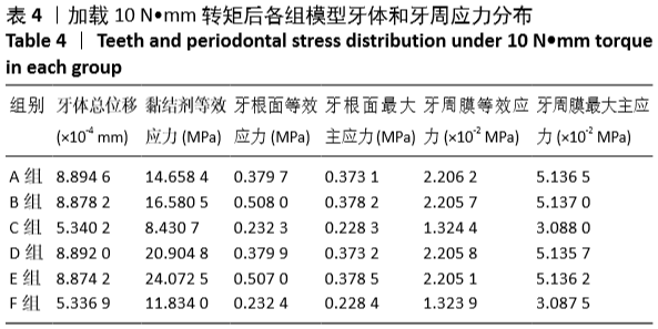
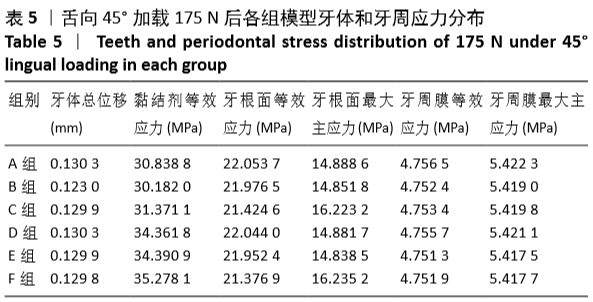
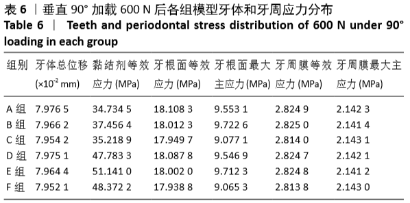
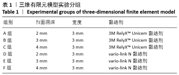
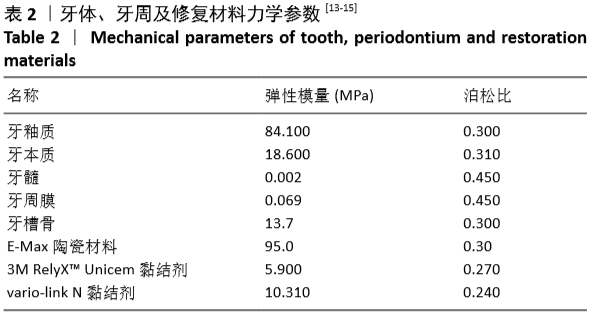
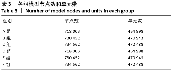
方法:利用Micro-CT扫描人下颌第三磨牙,采用医学建模软件mimics 20、逆向工程软件Geomagic Studio 2014、三维机械制图专用软件 NX 10建立不同黏结剂和不同洞深的近中-牙合-远中洞型E-Max瓷嵌体修复三维有限元模型:A组洞深2 mm,使用3M RelyX™ Unicem黏结剂;B组洞深3 mm,使用3M RelyX™ Unicem黏结剂;C组洞深4 mm,使用3M RelyX™ Unicem黏结剂;D组洞深2 mm,使用vario-link N黏结剂;E组洞深3 mm,使用vario-link N黏结剂;F组洞深4 mm,使用vario-link N黏结剂。应用有限元分析软件 ANSYS workbench 18.2进行网格划分,分析各模型在10 N•mm转矩、45°加载175 N和90°加载600 N时的应力分布状况。
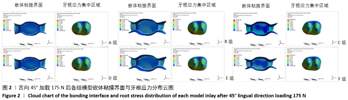
结果与结论:①加载10 N•mm转矩时,使用同一黏结剂的情况下,随着洞深度增加模型的牙体总位移、牙周膜等效应力均趋减小,洞深  3 mm时牙根面等效应力、黏结剂等效应力最大;在同一洞深下,使用vario-link N黏结剂模型的黏结剂等效应力和牙根面最大主应力较大;②舌向 45°加载175 N时,使用同一黏结剂的情况下,随着洞深度增加牙根面等效应力趋减小,洞深4 mm时牙体总位移、黏结剂等效应力最大,洞深2 mm时牙周膜等效应力、牙周膜最大主应力最大;在同一洞深下,使用3M RelyX™ Unicem黏结剂模型的牙根面最大主应力、牙根面等效应力、牙周膜等效应力较高,黏结剂等效应力较小;③舌向90°加载600 N时,使用同一黏结剂的情况下,随着洞深度增加模型的牙体总位移、牙周膜等效应力均趋减小,洞深3 mm时牙根面最大主应力与黏结剂等效应力最大,4 mm时牙周膜最大主应力最大;在同一洞深下,使用3M RelyX™ Unicem黏结剂模型的牙根面等效应力、牙体总位移、牙根面最大主应力和最大主应力较高,黏结剂等效应力较小;④结果表明,近中-牙合-远中洞型E-Max瓷嵌体修复三维有限元模型应力集中区域为牙根分叉区、嵌体边缘线、髓室顶、龈壁等,粘接界面和龈壁、根分叉区是应力集中和破坏的重点区域。

https://orcid.org/0000-0002-3185-6560 (张国梅) 

中国组织工程研究杂志出版内容重点:生物材料;骨生物材料; 口腔生物材料; 纳米材料; 缓释材料; 材料相容性;组织工程

关键词: 义齿, 材料, 三维有限元, 瓷嵌体, 黏结剂, 应力分布, 牙体缺损

Abstract: BACKGROUND: E-Max porcelain inlay has good aesthetic, bonding and mechanical properties, and has a broad application prospect in the field of tooth defect repair. 
OBJECTIVE: To build the model of mesio-occluso-distal cavity E-Max porcelain inlay with different adhesives and different depths of holes, and to explore the stress distribution and regional law of different data models.
METHODS: Micro-CT was used to scan the human mandibular third molar model. Medical modeling software mimics 20, reverse engineering software Geomagic Studio 2014, and three-dimensional mechanical drawing software NX 10 were utilized to construct the three-dimensional finite element models of mesio-occluso-distal cavity E-Max porcelain inlay with different adhesives and different depths of holes: Group A with a hole depth of 2 mm, using 3M RelyX™ Unicem adhesive; group B with a hole depth of 3 mm, using 3M RelyX™ Unicem adhesive; group C with a hole depth of 4 mm, using 3M RelyX™ Unicem adhesive; group D with a hole depth of 2 mm, using vario-link N adhesive; group E with a hole depth of 3 mm, using vario-link N adhesive; group F with a hole depth of 4 mm, using vario-link N adhesive. Finite element analysis software ANSYS workbench 18.2 was used for meshing. Stress distribution of each model at 10 N•mm torque, 45° loading 175 N and 90° loading 600 N was analyzed.  
RESULTS AND CONCLUSION: (1) After 10 N•mm torque loading, with the increase of cavity depth, total displacement of the teeth and the equivalent stress of the periodontal ligament decreased with the same adhesive; when the cavity depth was 3 mm, the root surface equivalent stress and the adhesive equivalent stress were largest. Under the same cavity depth, the equivalent stress and the maximum principal stress of the root surface were larger when using vario link N adhesive. (2) When 175 N was applied at 45° lingual direction and the same adhesive was used, the equivalent stress on the root surface decreased with the increase of the cavity depth. When the depth of the cavity was 4 mm, the total displacement of the tooth and the equivalent stress of the adhesive were largest. When the cavity depth was 2 mm, the equivalent stress and the maximum principal stress of periodontal ligament were largest. At the same cavity depth, the maximum principal stress, equivalent stress of root surface and equivalent stress of periodontal membrane of models using 3M RelyX™ T Unicem adhesive were higher and equivalent stress of the adhesive smaller than those of other models. (3) When 600 N was applied at 90° lingual direction and the same adhesive was used, with the increase of the cavity depth, the total displacement of the tooth and the equivalent stress of the periodontal membrane decreased. When the cavity depth was 3 mm, the maximum principal stress of the root surface and the equivalent stress of the adhesive were maximum; when the cavity depth was 4 mm, the maximum principal stress of periodontal ligament was largest. At the same cavity depth, the equivalent stress of the root surface, the total displacement of the tooth, the maximum principal stress and the maximum principal stress of the root surface were high, while the equivalent stress of the adhesive was small in models using 3M RelyX™ Unicem adhesive. (4) Results indicate that stress concentration areas are the root bifurcation area, the inlay edge line, the pulp chamber top, and the gingival wall; and key areas of stress concentration and destruction are the bonding interface, the gingival wall and the root bifurcation area in the three-dimensional finite element models of mesio-occluso-distal cavity E-Max porcelain inlay.

Key words: denture, material, three-dimensional finite element, porcelain inlay, adhesive, stress distribution, tooth defect

中图分类号: