中国组织工程研究 ›› 2020, Vol. 24 ›› Issue (24): 3857-3861.doi: 10.3969/j.issn.2095-4344.2706

• 骨与关节图像与影像 bone and joint imaging • 上一篇    下一篇

退变性腰椎滑脱患者坐-立位脊柱-骨盆矢状面序列的影像变化

刘  阳,徐宝山,许海委,黎  宁,姜洪丰,王  涛,刘  越   

  1. 天津市天津医院微创脊柱外科,天津市  300211
  • 收稿日期:2019-10-31 修回日期:2019-11-12 接受日期:2019-12-26 出版日期:2020-08-28 发布日期:2020-08-14
  • 通讯作者: 徐宝山,博士,硕士生导师,主任医师,天津市天津医院微创脊柱外科,天津市 300211
  • 作者简介:刘阳,男,1994年生,河南省平顶山市人,天津医科大学在读硕士,主要从事脊柱外科方面的研究。
  • 基金资助:

    国家自然科学基金资助项目(31670983);国家自然科学基金资助项目(31500781)

Imaging changes in spinal-pelvic sagittal alignment in sitting and standing positions in degenerative lumbar spondylolisthesis patients

Liu Yang, Xu Baoshan, Xu Haiwei, Li Ning, Jiang Hongfeng, Wang Tao, Liu Yue   

  1. Department of Minimally Invasive Spine Surgery, Tianjin Hospital, Tianjin 300211, China
  • Received:2019-10-31 Revised:2019-11-12 Accepted:2019-12-26 Online:2020-08-28 Published:2020-08-14
  • Contact: Xu Baoshan, MD, Master’s supervisor, Chief physician, Department of Minimally Invasive Spine Surgery, Tianjin Hospital, Tianjin 300211, China
  • About author:Liu Yang, Master candidate, Department of Minimally Invasive Spine Surgery, Tianjin Hospital, Tianjin 300211, China
  • Supported by:
    the National Natural Science Foundation of China, No. 31670983; the National Natural Science Foundation of China, No. 31500781

摘要:

文题释义:

退变性腰椎滑脱:是一类常见的老年退变性疾病,影像学上表现为在椎弓完整的情况下,一个椎体相对于另一个椎体向前或向后移位,好发于L4-5节段,临床上多表现为腰椎管狭窄症状,行走后症状加重,休息时减轻,保守治疗效果不佳时通常需要手术治疗。 

脊柱-骨盆矢状面序列:由脊柱和骨盆共同构成,在正常生理状态下,脊柱在矢状面呈“S”形,上承头颅,下连骨盆,有颈、胸、腰、骶4个生理弯曲,对于维持脊柱平衡具有重要意义。脊柱-骨盆矢状面序列的异常与退变性腰椎滑脱等疾病发生、发展相关,同时脊柱-骨盆矢状面序列受姿势的影响,当姿势变化时脊柱-骨盆矢状面序列也会发生相应改变。

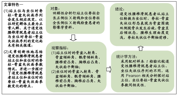
背景:脊柱-骨盆矢状面序列对于退变性腰椎滑脱的诊治十分重要,但是目前关于退变性腰椎滑脱患者脊柱-骨盆矢状面序列的研究局限在站立位体位,坐位下的脊柱-骨盆矢状面序列未见相关报道。

目的:分析退变性腰椎滑脱患者坐-立位脊柱-骨盆矢状位序列的影像学资料,探究退变性腰椎滑脱患者脊柱-骨盆矢状位序列从站立位到坐位的变化特点。

方法:纳入2019年3至9月天津市天津医院收治的44例退变性腰椎滑脱患者,其中男12例,女32例,年龄50-84岁,所有患者均拍摄站立位全脊柱X射线片和坐位全脊柱X射线片,通过院内影像归档与通信系统测量每例患者的骨盆入射角、骨盆倾斜角、骶骨倾斜角、腰椎前凸角、胸椎后凸角、矢状面平衡轴等参数,比较退变性腰椎滑脱患者站立位、坐位矢状位序列的不同,运用Pearson相关分析探讨站立位、坐位脊柱-骨盆矢状位参数间相关性。试验获得天津市天津医院伦理委员会批准。

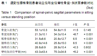
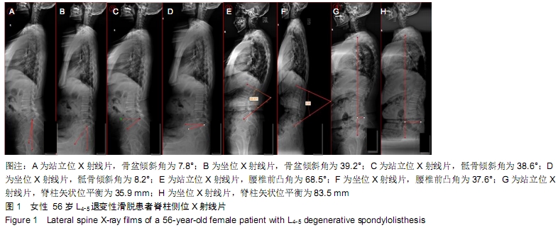
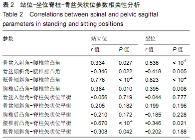
结果与结论:①由站立位转变为坐位时,44例退变性腰椎滑脱患者的骨盆倾斜角增大[(21.3±10.1)°,(34.0±10.4)°,P < 0.001]、骶骨倾斜角减小[(31.5±8.6)°,(20.8±12.7)°,P < 0.001]、腰椎前凸角减小[(40.9±14.6)°,(25.8±15.0)°,P < 0.001]、矢状面平衡轴增大[(43.0±43.4),(75.0±34.8)mm,P < 0.001],骨盆入射角与胸椎后凸角无明显改变(P > 0.05);②无论站位还是坐位,腰椎前凸角与其他5项参数均具有相关性(P < 0.05);由站位转换为坐位后,骶骨倾斜角与矢状位平衡参数矢状面平衡轴的相关性消失(P > 0.05),腰椎前凸角与矢状面平衡轴的相关性仍然存在(P < 0.05);③结果表明退变性腰椎滑脱患者从站位转换为坐位时,脊柱-骨盆矢状位形态表现为骨盆围绕双侧股骨头发生后旋,骨盆呈后倾状态,腰椎生理曲度变浅,脊柱矢状位平衡轴前移。

ORCID: 0000-0002-9690-3188(刘阳)

中国组织工程研究杂志出版内容重点:人工关节;骨植入物;脊柱骨折;内固定;数字化骨科;组织工程

关键词: 脊柱, 骨盆, 矢状位序列, 体位变化, 退变性腰椎滑脱, 站位, 坐位, 腰椎手术

Abstract:

BACKGROUND: Spinal-pelvic sagittal alignment is important for the diagnosis and treatment of degenerative lumbar spondylolisthesis. However, the current study of the spine-pelvic sagittal alignment in patients with degenerative lumbar spondylolisthesis is limited to the standing position. There is no relevant report on the spine-pelvic sagittal alignment under the sitting position.

OBJECTIVE: To analyze imaging data of sitting-standing spine-pelvic sagittal alignment in patients with degenerative lumbar spondylolisthesis, and to determine the sagittal alignment of spine change in degenerative lumbar spondylolisthesis patients from standing position to sitting position.

METHODS: Totally 44 patients with degenerative lumbar spondylolisthesis (12 males, 32 females; age, 50-84 years) were enrolled from Tianjin Hospital from March to September 2019. All patients took X-rays of the spine in standing and sitting positions. Through the hospital image archiving and communication system, spinal and pelvic parameters were measured, including pelvic incidence, pelvic tilt, sacral slope, lumbar lordosis, thoracic kyphosis, and sagittal vertical axis. The parameters were compared between standing posture and sitting posture. By using Pearson’s correlation test, differences of relationship between spinal and pelvic parameters in standing versus sitting position were discussed. This study was approved by the Ethics Committee of Tianjin Hospital.

RESULTS AND CONCLUSION: (1) When moving from standing to sitting position, in 44 degenerative lumbar spondylolisthesis patients, pelvic tilt increased [(21.3±10.1)°, (34.0±10.4)°, P < 0.001]; sacral slope decreased [(31.5±8.6)°, (20.8±12.7)°, P < 0.001]; lumbar lordosis reduced [(40.9±14.6)°, (25.8±15.0)°, P < 0.001]; sagittal vertical axis increased [(43.0±43.4), (75.0±34.8) mm, P < 0.001]; pelvic incidence and thoracic kyphosis did not significantly changed (P > 0.05). (2) Whether standing or sitting position, lumbar lordosis was correlated with other parameters (P < 0.05). When changing from standing to sitting position, the correlation between sacral slope and sagittal vertical axis disappeared (P > 0.05), but lumbar lordosis was also correlated with sagittal vertical axis (P < 0.05). (3) When the degenerative lumbar spondylolisthesis patients change from standing position to sitting position, the sagittal configuration of spine pelvis shows that the pelvis rotates back around the bilateral femoral heads; the pelvis shows a backward leaning state; the physiological curvature of lumbar spine becomes shallow; and the sagittal balance axis of spine moves forward.

Key words: spine, pelvis, sagittal alignment, position change, degenerative lumbar spondylolisthesis, standing position, sitting position, lumbar surgery

中图分类号: