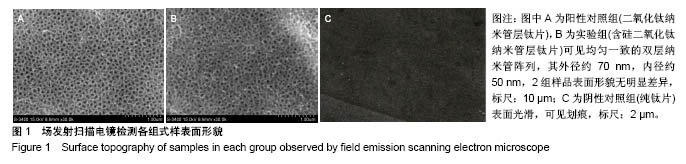
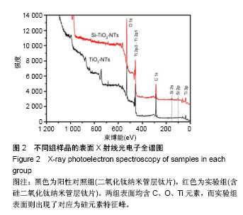
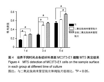
| [1] Cipriano AF, Miller C, Liu H. Anodic growth and biomedical applications of Ti02 nanotubes. J Biomed Nanotechnol. 2014;10(10):2977-3003.[2] Munirathinam B, Neelakantan L. Titania nanotubes from weak organic acid electrolyte: fabrication, characterization and oxide film properties.Mater Sci Eng C Mater Biol Appl. 2015;49:567-578.[3] Khan AF, Saleem M, Afzal A, et al. Bioactive behavior of silicon substituted calcium phosphate based bioceramics for bone regeneration. Mater Sci Eng C Mater Biol Appl. 2014;35:245-252.[4] Shi X, Nakagawa M, Kawachi G, et al. Surface modification of titanium by hydrothermal treatment in Mg-containing solution and early osteoblast responses. Mater Sci Mater Med. 2012;23(5):1281-1290.[5] Bauer S, Park J, Faltenbacher J, et al. Size selective behavior of mesenchymal stem cells on ZrO(2) and TiO(2) nanotube arrays. Integr Biol (Camb).2009;1(8-9):525-532.[6] Lou W, Dong Y, Zhang H, et al. Preparation and Characterization of Lanthanum-Incorporated Hydroxyapatite Coatings on Titanium Substrates. Int J Mol Sci. 2015;16(9):21070-21086.[7] Rao PJ, Pelletier MH, Walsh WR, Mobbs RJ. Spine interbody implants: material selection and modification, functionalization and bioactivation of surfaces to improve osseointegration. Orthop Surg.2014;6(2):81-89.[8] Matena J, Petersen S, Gieseke M, et al. SLM produced porous titanium implant improvements for enhanced vascularization and osteoblast seeding. Int J Mol Sci. 2015;16(4):7478-7492.[9] Chen WC, Chen YS, Ko CL, et al. Interaction of progenitor bone cells with different surface modifications of titanium implant. Mater Sci Eng C Mater Biol Appl.2014;37:305-313.[10] Kulkarui M, Mazare A, Gongadze E, et al. Titanium nanostruetnres for biomedical applications. Nanoteehnology. 2015;26(6):062002.[11] 杜庆华,曹均凯,董溪溪,等.镁黄长石浸提液诱导多潜能干细胞的成骨分化[J].中国组织工程研究,2015,19(14):2236-2242.[12] Zhang W, Jin Y, Qian S, et al. Vacuum extraction enhances rhPDGF-BB immobilization on nanotubes to improve implant osseointegration in ovariectomized rats. Nanomedicine. 2014;10(8):1809-1818.[13] Buttke TM, McCubrey JA, Owen TC. Use of an aqueous soluble tetrazolium/formazan assay to measure viability and proliferation of lymphokine-dependent cell lines. J Immunol Methods. 1993;157(1-2): 233-240.[14] Yamamoto O,Alvarez K,Kashiwaya Y,et al. Surface characterization and biological response of carbon-coated oxygen-diffused titanium having different topographical surfaces. J Mater Sci Mater Med. 2011;22(4):977-987.[15] Gong T, Xie J, Liao J, et al. Nanomaterials and bone regeneration. Bone Res.2015;3:15029.[16] Qiao Y, Zhang W, Tian P, et al. Stimulation of bone growth following zinc incorporation into biomaterials. Biomaterials. 2014;35(25):6882-6897.[17] Wang G, Li J, Zhang W, et al. Magnesium ion implantation on a micro/nanostructured titanium surface promotes its bioactivity and osteogenic differentiation function. Int J Nanomedicine. 2014;9:2387-2398.[18] Macdonald HM, Hardcastle AC, Jugdaohsingh R, et al. Dietary silicon interacts with oestrogen to influence bone health: evidence from the Aberdeen Prospective Osteoporosis Screening Study. Bone. 2012;50(3): 681-687.[19] Honda M, Kikushima K, Kawanobe Y, et al. Enhanced early osteogenic differentiation by silicon-substituted hydroxyapatite ceramics fabricated via ultrasonic spray pyrolysis route. J Mater Sci Mater Med. 2012;23(12): 2923-2932.[20] Rosemary FL, Suswillo, Behzad Javaheri, et al. Strain uses gap junctions to reverse stimulation of osteoblast proliferation by osteocytes. Cell Biochem Funct. 2017;35(1):56-65.[21] Minagar S, Li Y, Berndt CC, Wen C. The influence of titania-zirconia- zirconium titanate nanotube characteristics on osteoblast cell adhesion. Acta Biomater. 2015;12:281-289.[22] Sawada R, Kono K, Isama K, et al. Calcium-incorporated titanium surfaces influence the osteogenic differentiation of human mesenchymal stem cells. J Biomed Mater Res A. 2013;101(9):2573-2585.[23] Liu W, Arnaud Chaix, Magali Gary-Bobo, et al. Stealth Biocompatible Si-Based Nanoparticles for Biomedical Applications. Nanomaterials (Basel).2017;7(10):288.[24] Li J, Wei L, Sun J, et al. Effect of ionic products of dicalcium silicate coating on osteoblast differentiation and collagen production via TGF-beta1 pathway. J Biomater Appl. 2013;27(5):595-604.[25] Juliana AD, Mariana M, Celso AB, et al. Addition of Wollastonite Fibers to Calcium Phosphate Cement Increases Cell Viability and Stimulates Differentiation of Osteoblast-Like Cells. ScientificWorldJournal. 2017; 2017: 5260106.[26] Shahdadfar A, Fronsdal K, Haug T, et al. In vitro expansion of human mesenchymal stem cells: choice of serum is a determinant of cell proliferation, differentiation, gene expression, and transcriptome stability. Stem Cells. 2005;23(9):1357-1366.[27] Premnath P, Tan B, Venkatakrishnan K. Programming cell fate on bio-functionalized silicon. Colloids and surfaces B. Biointerfaces. 2015; 128:100-105.[28] Ling M, Huang PX, Islam S, et al. Epigenetic regulation of Runx2 transcription and osteoblast differentiation by nicotinamide phosphoribosyltransferase. Cell Biosci. 2017;7:27.[29] Ercan B, Webster TJ. The effect of biphasic electrical stimulation on osteoblast function at anodized nanotubular titanium surfaces. Biomaterials. 2010;31(13):3684-3693.[30] Wang C, Duan Y, Markovic B, et al. Phenotypic expression of bone-related genes in osteoblasts grown on calcium phosphate ceramics with different phase compositions. Biomaterials.2004;25(13):2507-2514.[31] Ducy P, Zhang R, Geoffroy V, et al. Osf2/Cbfa1: a transcriptional activator of osteoblast differentiation. Cell. 1997;89(5):747-754.[32] Geoffroy V, Kneissel M, Fournier B, et al. High bone resorption in adult aging transgenic mice overexpressing cbfa1/runx2 in cells of the osteoblastic lineage. Mol Cell Biol.2002;22(17):6222-6233.[33] Franceschi RT, Xiao G, Jiang D, et al. Multiple signaling pathways converge on the Cbfa1/Runx2 transcription factor to regulate osteoblast differentiation. Connect Tissue Res. 2003;44(Suppl 1):109-116. |
.jpg)


.jpg)
.jpg)