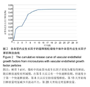
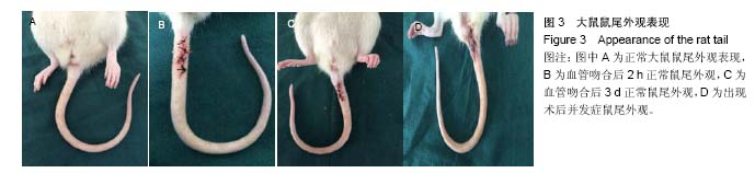
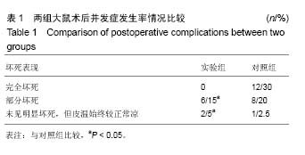
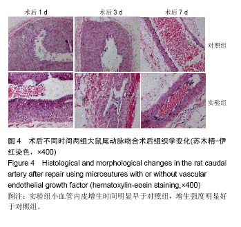
| [1]Zhang L,Si T,Fischer AJ,et al.Coaxial Electrospray of Ranibizumab- Loaded Microparticles for Sustained Release of Anti-VEGF Therapies. PLoS One.2015;10(8):e0135608. [2]Park K.Delivery of definable numbers of PLGA microparticles within embryoid bodies.J Control Release,.2013;168(1):103. [3]Herrán E,Ruiz-Ortega JÁ,Aristieta A,et al.In vivo administration of VEGF- and GDNF-releasing biodegradable polymeric microspheres in a severe lesion model of Parkinson's disease.Eur J Pharm Biopharm. 2013;85(3 Pt B):1183-1190. [4]Simón-Yarza T,Tamayo E,Benavides C,et al.Functional benefits of PLGA particulates carrying VEGF and CoQ10 in an animal of myocardial ischemia.Int J Pharm.2013;454(2):784-790. [5]White LJ,Kirby GT,Cox HC,et al.Accelerating protein release from microparticles for regenerative medicine applications.Mater Sci Eng C Mater Biol Appl.2013;33(5):2578-2583. [6]Dhanda DS,Tyagi P,Mirvish SS,et al.Supercritical fluid technology based large porous celecoxib-PLGA microparticles do not induce pulmonary fibrosis and sustain drug delivery and efficacy for several weeks following a single dose.J Control Release.2013;168(3):239-250. [7]Qutachi O,Shakesheff KM,Buttery LD,et al.Delivery of definable number of drug or growth factor loaded poly(DL-lactic acid-co-glycolic acid microparticles within human embryonic stem cell derived aggregates.J Control Release.2013;168(1):18-27.[8]Bible E,Qutachi O,Chau DY,et al.Neo-vascularization of the stroke cavity by implantation of human neural stem cells on VEGF-releasing PLGA microparticles.Biomaterials.2012;33(30):7435-7446.[9]Simón-Yarza T,Formiga FR,Tamayo E,et al.PEGylated-PLGA microparticles containing VEGF for long term drug delivery.Int J Pharm.2013;440(1):13-18. [10]Xu H,Nguyen KT,Brilakis ES,et al.Enhanced endothelialization of a new stent polymer through surface enhancement and incorporation of growth factor-delivering microparticles.J Cardiovasc Transl Res. 2012;5(4):519-527. [11]Formiga FR,Pelacho B,Garbayo E,et al.Sustained release of VEGF through PLGA microparticles improves vasculogenesis and tissue remodeling in an acute myocardial ischemia-reperfusion model.J Control Release.2010;147(1):30-37. [12]Shahani K,Swaminathan SK,Freeman D,et al.Injectable sustained release microparticles of curcumin: a new concept for cancer chemoprevention.Cancer Res.2010;70(11):4443-4452. [13]Jay SM,Shepherd BR,Bertram JP,et al.Engineering of multifunctional gels integrating highly efficient growth factor delivery with endothelial cell transplantation.FASEB J.2008;22(8):2949-2956. [14]Amrite AC,Ayalasomayajula SP,Cheruvu NP,et al.Signgle periocular injection of celecoxib-PLGA microparticles inhibits diabetes-induced elevations in retinal PGE2, VEGF, and vascular leakage.Invest Ophthalmol Vis Sci.2006;47(3):1149-1160.[15]Bandi N,Ayalasomayajula SP,Dhanda DS,et al.Intratracheal budesonide-poly(lactide-co-glycolide) microparticles reduce oxidative stress,VEGFexpression, and vascular leakage in a benzo(a)pyrene-fed mouse model.J Pharm Pharmacol.2005;57(7):851-860.[16]Chen Y,Luo X,Xiao J,et al.Experimental study on polylactic glycolate acid microparticles with releasing-slowly vascular endothelial growth factor.Zhongguo Xiu Fu Chong Jian Wai Ke Za Zhi. 2005;19(4):262-263. [17]Ayalasomayajula SP,Kompella UB.Subconjunctivally administered celecoxib-PLGA microparticles sustain retinal drug levels and alleviate diabetes-induced oxidative stress in a rat model.Eur J Pharmacol. 2005;511(2-3):191-198.[18]陈扬,肖建德,李振宇,等.血管内皮生长因子缓释微粒的合成与制备[J].中国骨肿瘤骨病杂志,2006,3(3):150-151.[19]沈素祥,美小涛,张绍东,等.rhBMP-2/无定形磷酸钙纳米缓释微粒成骨活性的实验研究[J].生物医学工程与临床杂志, 2008,3(12):175-178.[20]潘兴纳,李亚雄,蒋立虹.组织工程血管支架材料的研究与进展[J].中国组织工程研究,2016,20(34):5149-5154.[21]Gaitskell K,Martinek I,Bryant A,et al.Angiogenesis inhibitors for the treatment of ovarian cancer. Cochrane Database Syst Rev.2011;(9): CD007930.[22]Kroep JR,Nortier JWR.The role of bevacizumab in advanced epithelial ovarian cancer.Curr Pharm Des.2012;18(25):3775-3783.[23]Yamazaki Y,Morita T.Molecular and functional diversity of vascular endothelial growth factors. Mol Divers. 2006;10(4):515-527.[24]Roy H,Bhardwaj S,Ylä-Herttuala S. Biology of vascular endothelial growth factors.FEBS Lett. 2006;580(12):2879-2887.[25]Jackson MR,Carney EW,Lye SJ,et al.Localization of two angiogenic growth factors (PDECGF and VEGF) in human placentae throughout gestation.Placenta.1994;15(4):341-353.[26]Wei MH,Popescu NC,Lerman MI,et al.Localization of the human vascular endothelial growth factor gene, VEGF, at chromosome 6p12.Hum Genet.1996;97(6):794–797.[27]Vincenti V,Cassano C,Rocchi M,et al.Assignment of the vascular endothelial growth factor gene to human chromosome 6p21.3. Circulation.1996;93(8):1493-1495.[28]Hsieh YY,Chang CC,Tsai FJ,et al.Tallele for VEGF-460 gene polymorphism at 5’-untranslated region is associated with higher susceptibility of leiomyoma.Biochem Genet.2008;46(5-6):356-361.[29]Feng Y,Lin X,Zhou S,et al.The associations between the polymorphisms of the ER-α gene and the risk of uterine leiomyoma(ULM).Tumour Biol.2013;34(5):3077-3082.[30]Hsieh YY,Chang CC,Tsai FJ,et al.Estrogen receptor thymine-adenine dinucleotide repeat polymorphism is associated with susceptibility to leiomyoma.Fertil Steril.2003;79(1):96-99.[31]Yaghmaei M,Salimi S,Namazi L,et al.Association of XRCC1 Arg399GIn and Tp53 Arg72Pro polymorphisms and increased risk of uterine leiomyoma-a case-control study.Genet Mol Biol. 2015;38(4): 444-449.[32]Wang F,Chen J,Wang L,et al.CYP1A1 genetic polymorphisms and uterine leiomyoma risk: a meta-analysis.Int J Clin Exp Med. 2015;8(3): 3590-3594.[33]Feng Y,Zhao X,Zhou C,et al.The associations between the Val158Met in the catechol-O-methyltransferase(COMT) gene and the risk of uterine leiomyoma(ULM). Gene.2013;529(2): 296-299.[34]Salimi S,Hajizadeh A,Khodamian M,et al.Age-dependent association of MDM2 promoter polymorphisms and uterine leiomyoma in South-East Iran: A preliminary report.J Obstet Gynaecol Res.2015; 41(5):729-734.[35]Stoner M,Wang F,Wormke M,et al.Inhibition of vascular endothelial growth factor expression in HEC1A endometrial cancer cells through interactions of estrogen receptor alpha and Sp3 proteins.J Biol Chem. 2000;275(30):22769-22779.[36]Adams J,Carder PJ,Downey S,et al.Vascular endothelial growth factor(VEGF) in breast cancer: comparison of plasma, serum, and tissue VEGF and microvessel density and effects of tamoxifen.Cancer Res.2000;60(11):2898-2905.[37]Chao Y,Li CP,Chau GY,et al.Prognostic significance of vascular endothelial growth factor, basic fibroblast growth factor, and angiogenin in patients with resectable hepatocellular carcinoma after surgery.Ann Surg Oncol.2003;10(4):355-362. |
.jpg)
.jpg)
.jpg)