| [1]孙安科,陈文弦,崔鹏程,等.同种异体工程化软骨的构建和修复甲状软骨缺损的实验研究[J].中华耳鼻咽喉科杂志, 2001,36(4):278-280.
[2]李鹏程,程代薇,徐福,等.组织工程肌腱种子细胞的比较研究[J].组织工程与重建外科杂志,2006,2(3):134-136.
[3]孙安科,陈文弦,崔鹏程,等.以PGA 为三维支架同种异体工程化软骨的构建[J].解放军医学杂志, 2001,26(10): 748-749.
[4]Yeo MG,Lee H,Kim GH.Three-dimensional Hierarchical composite Scaffolds Consisting of Polycaprolactone, β-Tricalcium Phosphate, and Collagen Nanofibers: Fabrication, Physical Properties, and In vitro Cell Activity for Bone Tissue Regeneration. Biomacromolecules.2011;12(2):501-510.
[5]周栋,胡平,汪钢,等.聚羟基丁酸及其共聚物在体内组织相容性和降解率的实验研究[J].武警医学, 2003,14(4): 210-212.
[6]欧阳少平,丘远征,卢晓云,等.代谢工程技术调控3一羟基丁酸与3一羟基己酸共聚酯PHBHHx的微生物合成[J].生物加工过程,2003,1(1):60-65.
[7]Teixeira CR,Rahal SC,Volpi RS,et al.Tibial segmental bone defect treated with bone plate and cage filled with either xenogeneic composite or autologous cortical bone graft.An experimental study in sheep.Vet Comp Orthop Traumatol.2007;20(4):269-276.
[8]郭霄飞,张永红,杜美丽,等.羟基丁酸与羟基辛酸共聚物一体化骨软骨组织工程支架的制备及性能[J].中国组织工程研究,2012,16(16):2865-2868.
[9]李鹏程,程代薇,徐福,等.组织工程肌腱种子细胞的比较研究[J].组织工程与重建外科杂志,2006,2(3):134-136.
[10]胡晓洁,王敏,柴岗,等.组织工程技术构建兔角膜基质组织的实验研究[J].中华眼科杂志,2004,40(8):517-521.
[11]杨光辉,崔磊,刘伟,等.利用聚羟基乙酸构建组织工程皮肤的实验研究[J].中华实验外科杂志, 2003,20(l1): 984-985.
[12]田敏,王贻宁,陈新明,等.PGA无纺网与PDLCs的3D共培养物在裸鼠体内的生长观察[J].口腔医学研究, 2005, 21(2):116-118.
[13]孙安科,李万同,刘松波,等.聚羟基烷酸酯聚合物负载软骨细胞修复同种异体喉软骨缺损[J].中国组织工程研究, 2013,17(41):7181-7187.
[14]Gilpin DA,Weidenbecher MS,Dennis JE.Scaffold-free tissue-engineered cartilage implants for laryngotracheal reconstruction. Laryngoscope. 2010; 120(3):612-617.
[15]Ye C,Hu P,Ma MX,et al.PHB/PHBHHx scaffolds and human adipose-derived stem cells for cartilage tissue engineering.Aterials.2009;30(26):4401-4406.
[16]郭霄飞,张永红,杜美丽,等.羟基丁酸与羟基辛酸共聚物一体化骨软骨组织工程支架的制备及性能[J].中国组织工程研究,2012,16(16):2865-2868.
[17]Wang Y,Bian YZ,Wu Q,et al. Evaluation of three-dimensional scaffolds prepared from poly(3-hydroxybutyrate-co-3-hydroxyhexanoate)for growth of allogeneic chondrocytes for cartilage repair in rabbits.Biomaterials.2008;29(19):2858-2868.
[18]Sterodimas A,de Faria J.Human auricular tissue engineering in an immunocompetent animal model.Aesthet Surg J.2013;33(2):283-289.
[19]Koch H,Tomaselli F,Pierer G,et al.Thoracic wall reconstruction using both portions of the latissimus dorsi previously divided in the course of posterolateral thoracotomy. Eur J Cardiothorac Surg, 2002;21(5): 874-878.
[20]吴延平,吴方.聚羟基烷酸酯聚合物负载软骨细胞修复同种异体喉软骨缺损[J].中国组织工程研究, 2015,19(38): 6140-6144.
[21]孙安科,李万同,孟庆延,等.带蒂肌筋膜瓣充填与包裹构建喉支架形态组织工程软骨[J].中华耳鼻咽喉头颈外科杂志,2011,46(12):1019-1023.
[22]Nishimoto S,Fukuda K,Kawai K,et al.Supplementation of bone marrow aspirate-derived platelet-rich plasma for treating radiation-induced ulcer after cardiac fluoroscopic procedures: A preliminary report.Indian J Plast Surg.2012;45(1):109-114.
[23]张雷,于洪波,矫晓昆,等.应用液态及凝胶态生物载体材料负载同种异体软骨细胞修复全厚关节软骨缺损[J].中国临床康复,2006,10(45):190-193.
[24]Stangenberg L,Schaefer DJ,Buettner O,et al. Differentiation of osteo-blasts in three-dimensional culture in processed cancellous bone ma-trix: quantitative analysis of gene expression based onreal-time reverse transcription-polymerase chain reaction.Tissue Eng.2005;11:855-864.
[25]Rasmusson I, Ringdén O, Sundberg B, et al. Mesenchymal stem ceils inhibit lymphocyte proliferation by mitogens and alloantigens by different mechanisma.Exp Cell Res. 2005;305(1):33-41.
[26]Jakcb RP,Franz T,Gautier E,et al.Autologous osteoehondral grafting in the knee: Indication, results, and reflection.Clin Orthop Ralet Res. 2002;(401):170-184.
[27]Koulalis D,Di Benedetto P,Citak M,et al.Comparative study of navigated versus freehand osteochondral graft transplantation of the knee.Am J Sports Med. 2009;37(4):803-807.
[28]Bhumiratana S,Eton RE,Oungoulian SR,et al. Large,stratified, and mechanically functional human cartilage grown in vitro by mesenchymal condensation. Proc Natl Acad Sci US A.2014;111(19):6940-6945.
[29]Park JS,Yang HN,Woo DG,et al.Chondrogenesis of human mesenchymal stem cells in fibrin constructs evaluated in vitro and in nude mouse and rabbit defects models. Biomaterials.2011;32(6):1495-1507.
[30]Wei B,Jin C,Xu Y,et al.Effect of bone marrow mesenchymal stem cells-derived extracellular matrix scaffold on chondrogenic differentiation of marrow clot after microfracture of bone marrow stimulation in vitro. Zhongguo Xiu Fu Chong Jian Wai Ke Za Zhi. 2013; 27(4):464-474.
[31]孙安科,胡平,李万同,等.组织工程PHBHH多孔材料喉支架的制备与细胞相容性研究[J].听力学及言语疾病杂志, 2011,19(6):558-561.
[32]Maccario R,Podest M,Moretta A,et al.Interaction of human mesenchymal stem cells with cells involved in alloantigen-specific immune response favors the differentiation of CD 4+T-cell subsets expressing a regulatory/suppressive phenutype.Haematologica. 2005;90(4):516-525.
[33]闫磊,李思源,蔡晓玲.SD新西兰白兔骨髓间充质干细胞分离培养及其生物学特性鉴定[J].实用医学杂志, 2008, 24(17):2945-2947.
[34]Tang QO,Carasco CF,Gamie Z,et al.Preclinical and clinical data for the use of mesenchymal stem cells in articular cartilage tissue engineering. Expert Opin Biol Ther. 2012;12(10):1361-1382.
[35]Xie J,Han Z,Naito M,et al.Articular cartilage tissue engineering based on a mechano-active scaffold made of poly (L-lactide- co-epsilon-caprolactone): in vivo performance in adult rabbits.J Biomed Mater Res B Appl Biomater.2010;94(1):80-88.
[36]Crawford DC,Heveran CM,Cannon WD,et al.An autologous carti-lage tissue implant NeoCart for treatment of grade III chondral injury to the distal femur: prospective clinical safety trial at 2 years.Am J Sports Med.2009;37(7):1334-1343.
[37]Brix MO,Stelzeneder D,Trattnig S,et al.Cartilage repair of the knee with Hyalograft C: magnetic resonance imaging assessment of the glycosaminoglycan content at midterm. Int Orthop.2013;37(1):39-43.
[38]孙安科,李万同,刘松波等.聚羟基烷酸酯聚合物负载软骨细胞修复同种异体喉软骨缺损[J].中国组织工程研究, 2013,17(41):7181-7187.
[39]孙安科,胡平,李万同,等.组织工程PHBHH多孔材料喉支架的制备与细胞相容性研究[J].听力学及言语疾病杂志, 2011,19(6):558-561.
[40]王淑芳,郭天瑛,杨超,等.微生物合成的β-羟基丁酸酯与β-羟基已酸酯共聚物/聚大酸共混材料(PHBHHx/PLA)的力学性能与生物降解性研究[J].离子交换与吸附, 2006, 22(1):1-8.
[41]刘清宇,王富友,杨柳.关节软骨组织工程支架的研究进展[J].中国修复重建外科杂志,2012,26(10):1247-1250.
[42]Eslahi N,Abdorahim M,Simchi AA.Smart Polymeric Hydrogels for Cartilage Tissue Engineering: A Review on the Chemistry and Biological Functions. Biomacromolecules.2016.
[43]Liu W,Wang D,Huang J,et al.Low-temperature deposition manufacturing: A novel and promising rapid prototyping technology for the fabrication of tissue-engineered scaffold.Mater Sci Eng C Mater Biol Appl.2017;70(Pt 2):976-982.
[44]Wang J,Sun B,Tian L,et al.Evaluation of the potential of rhTGF- β3 encapsulated P(LLA-CL)/collagen nanofibers for tracheal cartilage regeneration using mesenchymal stems cells derived from Wharton's jelly of human umbilical cord.Mater Sci Eng C Mater Biol Appl.2017;70(Pt 1):637-645.
[45]Pouran B,Arbabi V,Weinans H,et al.Application of multiphysics models to efficient design of experiments of solute transport across articular cartilage.Comput Biol Med.2016;78:91-96.
[46]Bardsley K,Kwarciak A,Freeman C,et al.Repair of bone defects in vivo using tissue engineered hypertrophic cartilage grafts produced from nasal chondrocytes. Biomaterials.2016;112:313-323.
[47]Song K,Li W,Wang H,et al.Development and fabrication of a two-layer tissue engineered osteochondral composite using hybrid hydrogel-cancellous bone scaffolds in a spinner flask. Biomed Mater.2016;11(6):065002.
[48]Moshtagh PR,Pouran B,Korthagen NM,et al. Guidelines for an optimized indentation protocol for measurement of cartilage stiffness: The effects of spatial variation and indentation parameters.J Biomech.2016;49(14):3602-3607. |
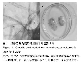
.jpg)
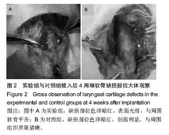
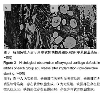
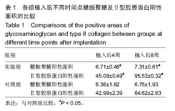
.jpg)