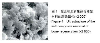
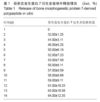
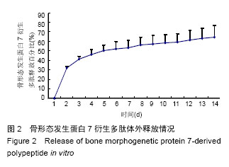
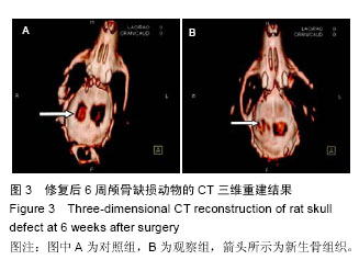
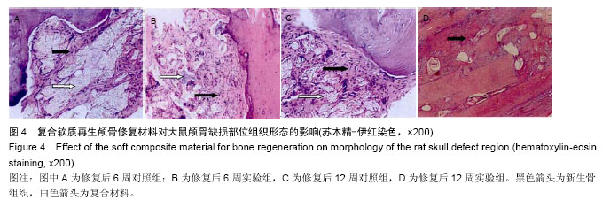
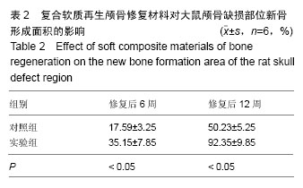
| [1] 王冠聪.具有二级三维网络结构的壳聚糖/羟基磷灰石骨组织工程复合支架材料的构建及其生物性能研究[D].济南:山东大学,2012. [2] 郭晓东,王明波,黎维勇,等.BMP2活性肽/PLGA复式微球及仿生支架材料的研制[C]//第九届中国南方骨质疏松论坛暨江西省骨质疏松学术会论文集.2013:198.[3] 李丹.PLGA/羟基磷灰石复合支架的制备及其用于骨修复的研究[D].杭州:浙江大学,2011.[4] 杨大志,罗惠莉,易伟宏,等.骨形态发生蛋白-2/胶原/掺锶羟基磷灰石材料修复颅骨缺损[J].中华实验外科杂志, 012,29(7):1250-1253. [5] 李均荣.羟基磷灰石/聚乳酸—聚羟乙酸/2型骨形态发生蛋白(HA-PLGA-BMP-2)颅骨修复材料的组织相容性及成骨性研究[D].张家口:河北北方学院,2013.[6] 张艳红.羟基磷灰石/丝素复合支架负载BMP-2的制备及其生物相容性研究[D].杭州:浙江大学,2011.[7] Cao N, Dong J, Wang Q, et al. An experimental bone defect healing with hydroxyapatite coating plasma sprayed on carbon/carbon composite implants. Surf Coat Technol. 2010;205(4):1150-1156.[8] Zhu W, Lu W, Zhang XJ, et al. Nano-hydroxyapatite/ fibrin glue/recombinant human osteogenic protein-1 artificial bone for repair of bone defect in an animal model. Micro Nano Lett. 2012;7(5):467-471.[9] Dong JL, Li LX, Mu WD, et al. Bone Regeneration with BMP-2 Gene-modified Mesenchymal Stem Cells Seeded on Nano-hydroxyapatite/Collagen/ Poly(L-Lactic Acid) Scaffolds. J Bioact Compat Polym. 2010;25(6):547-566.[10] 吴斌.胶原矿化仿生骨联合骨形态发生蛋白2相关多肽促进骨再生的实验研究[D].武汉:华中科技大学,2009.[11] 赵峰,尹玉姬,姚康德,等.壳聚糖-明胶网络/羟基磷灰石复合材料支架的研究--成骨细胞培养[J].中国修复重建外科杂志,2002,16(2):130-133.[12] 杨为中,周大利,尹光福,等.BMP/α-磷酸三钙生物活性骨水泥的BMP释放动力学及成骨性能研究[J].无机材料学报,2004,19(6):1359-1366.[13] 熊莹帆,邱建华,楚胜华,等.不同颅骨成型材料的选择及评价[J].中华临床医师杂志(电子版), 2011,5(17): 5119-5121.[14] Nitzsche H, Lochmann A, Metz H, et al. Fabrication and characterization of a biomimetic composite scaffold for bone defect repair. J Biomed Mater Res A. 2010;94(1):298-307.[15] Jung UW, Song KY, Kim CS, et al. Effects of a chitosan membrane coated with polylactic and polyglycolic acid on bone regeneration in a rat calvarial defect. Biomed Mater. 2007;2(3):S101-105.[16] Zhang X, Zhu L, Lv H, et al. Repair of rabbit femoral condyle bone defects with injectable nanohydroxyapatite/chitosan composites. J Mater Sci Mater Med. 2012;23(8):1941-1949.[17] 徐建军,丁华山,瞿鸿义,等.计算机三维重建EH复合型颅骨预制体修复额颞顶大面积颅骨缺损[J].中国临床神经外科杂志,2006,11(12):748-749.[18] 章浩伟,周思远,刘颖,等.基于增材制造技术修复缺损颅骨的研究和应用[J].生物医学工程学进展, 2014,35(4): 219-222.[19] Dinarvand P, Seyedjafari E, Shafiee A, et al. New approach to bone tissue engineering: simultaneous application of hydroxyapatite and bioactive glass coated on a poly(L-lactic acid) scaffold. ACS Appl Mater Interfaces. 2011;3(11):4518-4524.[20] Nakasa T, Ishida O, Sunagawa T, et al. Feasibility of prefabricated vascularized bone graft using the combination of FGF-2 and vascular bundle implantation within hydroxyapatite for osteointegration. J Biomed Mater Res A. 2008;85(4):1090-1095.[21] 王新.纳米羟基磷灰石-壳聚糖骨组织工程支架的研究[D].北京:中国协和医科大学,2007.[22] 杨耀武,毛天球,侯锐,等.鸵鸟骨转化羟基磷灰石支架复合骨膜修复颅骨极限缺损实验研究[J].实用口腔医学杂志, 2007,23(3):317-320.[23] 徐建军,丁华山,瞿鸿义,等.颅骨成形术三种不同材料的临床分折[J].中国临床神经外科杂志,2003,8(1):67-68.[24] 叶迅,赵元立,朱琳等.颅骨修补材料钛金属表面梯度生物活性涂层的生物相容性研究[J].北京医学, 2008,30(1): 8-12.[25] Florczyk SJ, Leung M, Li Z, et al. Evaluation of three-dimensional porous chitosan-alginate scaffolds in rat calvarial defects for bone regeneration applications. J Biomed Mater Res A. 2013;101(10): 2974-2983.[26] 孙春明,崔岗,周岱.EH复合材料人工颅骨的生物力学研究[J].苏州医学院学报,1999,19(9):969-970.[27] 唐晓军.天然羟基磷灰石/壳聚糖复合材料生物学特性的实验研究[D].北京:中国协和医科大学,2006.[28] 施恒军,曹胜武.医用树脂和羟基磷灰石复合材料在修补大面积颅骨缺损中的应用体会(附42例报告)[J].实用临床医药杂志,2011,15(19):153-154.[29] Bi L, Jung S, Day D, et al. Evaluation of bone regeneration, angiogenesis, and hydroxyapatite conversion in critical-sized rat calvarial defects implanted with bioactive glass scaffolds. J Biomed Mater Res A. 2012;100(12):3267-3275.[30] 田轶.颗粒状羟基磷灰石与纤维蛋白凝胶复合人工骨的可塑性和生物相容性的实验研究[D].大连:大连医科大学, 2007.[31] Ma X, Wang Y, Guo H, et al. Nano-hydroxyapatite/ chitosan sponge-like biocomposite for repairing of rat calvarial critical-sized bone defect. J Bioact Compat Polym. 2011;26(4): 335-346.[32] Wang YY, Shi R, Gong P, et al. Bioelectric effect of a chitosan bioelectret membrane on bone regeneration in rabbit cranial defects. J Bioact Compat Polym. 2012; 27(2):122-132.[33] 傅德皓.骨形态发生蛋白-2诱导成骨过程中血管内皮生长因子的表达及意义[D].武汉:华中科技大学,2006.[34] Datta P, Ghosh P, Ghosh K, et al. In vitro ALP and osteocalcin gene expression analysis and in vivo biocompatibility of N-methylene phosphonic chitosan nanofibers for bone regeneration. J Biomed Nanotechnol. 2013;9(5):870-879.[35] Cui ZW, Wright LD, Guzzo R, et al. Poly(D-lactide)/ poly(caprolactone) nanofiber thermogelling chitosan gel composite scaffolds for osteochondral tissue regeneration in a rat model. J Bioact Compat Polym. 2013;28(2):115-125.[36] Fricain JC, Schlaubitz S, Le Visage C, et al. A nano-hydroxyapatite--pullulan/dextran polysaccharide composite macroporous material for bone tissue engineering. Biomaterials. 2013;34(12):2947-2959. [37] Lü YM, Cheng LM, Pei GX, et al. Experimental study of repairing femoral bone defects with nHA/RHLC/PLA scaffold composite with endothelial cells and osteoblasts in canines. Zhonghua Yi Xue Za Zhi. 2013;93(17):1335-1340. [38] Oliveira JM, Rodrigues MT, Silva SS, et al. Novel hydroxyapatite/chitosan bilayered scaffold for osteochondral tissue-engineering applications: Scaffold design and its performance when seeded with goat bone marrow stromal cells. Biomaterials. 2006; 27(36):6123-6137. [39] Kim SS, Sun Park M, Jeon O, et al. Poly(lactide-co-glycolide)/hydroxyapatite composite scaffolds for bone tissue engineering. Biomaterials. 2006;27(8):1399-1409. [40] Laschke MW, Strohe A, Menger MD, et al. In vitro and in vivo evaluation of a novel nanosize hydroxyapatite particles/poly(ester-urethane) composite scaffold for bone tissue engineering. Acta Biomater. 2010;6(6):2020-2027. [41] Xue D, Zheng Q, Zong C, et al. Osteochondral repair using porous poly(lactide-co-glycolide)/ nano-hydroxyapatite hybrid scaffolds with undifferentiated mesenchymal stem cells in a rat model. J Biomed Mater Res A. 2010;94(1):259-270. [42] Zhang W, Walboomers XF, van Osch GJ, et al. Hard tissue formation in a porous HA/TCP ceramic scaffold loaded with stromal cells derived from dental pulp and bone marrow. Tissue Eng Part A. 2008;14(2):285-294.[43] 车向新.诱导型人工骨材料修复兔颅骨缺损的实验研究[D].南昌:南昌大学,2008.[44] 胡鹏.犬甲状软骨缺损应用骨形态发生蛋白材料修复的实验研究[D].昆明:昆明医学院,2001. |
.jpg)
.jpg)
.jpg)