| [1] Ginsberg HN,Elam MB,Lovato LC,et al.Effects of combination lipid therapy in type 2 diabetes mellitus.N Engl J Med. 2010; 362(17):1563-1574.
[2] Kuro-o M,Matsumura Y,Ailawa H,et al.Mutation of the mouse klotho gene leads to a syndrome resembling ageing. Nature. 1997;390(6655):45-51.
[3] Kumsu H, Yamamoto M, Clark JD, et al. Suppression of aging in mice by the hormone Klotho. Science. 2005;309(5742):1829-1833.
[4] Aizawa H,Saito Y,Nakamura T,et al.Downregulation of the klotho gene in the kidney under sustained circulatory stress in rats.Biochem Biophys Res Commun. 1998;249(3):865-871.
[5] Arking DE,Becker DM,Yanek LR,et al.KLOTHO allele status and risk of early-onset occult coronary artery disease. Am J Hum Genet. 2003;72(5):1154-1161.
[6] Matsumura Y, Aizawa H, Shiraki-Iida T, et al. Identification of the human klotho gene and its two transcripts encoding membrane and secreted klotho protein.Biochem Biophys Res Commun. 1998;242(3): 626-630.
[7] Raekleum N, Rackietan ML, Nadkami MR. Studies on Diabetogenic action of astreptozotocin (NSC-37917). Cancer Chemother Rep. 1993;29:91.
[8] 王虎林,司良毅,孙兰英,等.Klotho基因G-395A多态性与动脉硬化的相关性研究[J].中华老年心脑血管病杂志,2008,10(2): 96-98.
[9] Takeshita K,Yamamoto K,Ito M,et al. Increased expression of plasminogen activator inhibitor-1 wirh fibrin deposition in a murine model of aging,”Klotho” mouse. Semin Thromb Hemost. 2002;28(6):545-554.
[10] Biddinger SB,Hernandez-Ono A,Rask-Maden C,et al. Hepatic insulin resistance is sufficient to produce dyslipidemia and susceptibility to atherosclerosis. Cell Metab. 2008;7(2):125-134.
[11] Price MJ, Shah PK. New strategies in managing and preventing atherosclerosis: focus on HDL. Rev Cardiovasc Med. 2002;3(3):129-137.
[12] 房晨鹏,刘怡欣,田鹏,等.Klotho基因多态性与低密度脂蛋白水平的关系[J].现代预防医学, 2011,38(14):2818-2820.
[13] Arking DE, Becker DM, Yanek LR, et al. KLOTHO allele status and the risk of early-onset occult coronary artery disease. Am H Hum Genet. 2003;72:1154-1161.
[14] Arking DE,Atzmon G,Arking A,et al.Association between a funcitional variant of the KLOTHO gene and high-density lipoprotein cholesterol,blood pressure,stroke,and longevity. Circ Res. 2005;96:412-418.
[15] Wang HL, Xu Q, Wu Z, et al. A potential regulatory single nucleotide polymorphism in the promoter of the Klotho gene may be associated with essential hypertension in the Chinese Han population. Clinica Chimica Acta. 2010;411: 386-390.
[16] Shu L, Chen M, Wen M, et al. Study of klotho gene transfer for the protective effect of the coronary of diabetic rats. Pak J Pharm Sci. 2014;27(6 Suppl):2095-2099.
[17] Navarro-González JF, Donate-Correa J, de Fuentes M, et al. Reduced Klotho is associated with the presence and severity of coronary artery disease. Heart. 2014;100(1):34-40.
[18] Ko GJ, Lee YM, Lee EA, et al. The association of Klotho gene polymorphism with the mortality of patients on maintenance dialysis. Clin Nephrol. 2013;80(4):263-269.
[19] Tangri N, Alam A, Wooten EC, et al. Lack of association of Klotho gene variants with valvular and vascular calcification in Caucasians: a candidate gene study of the Framingham Offspring Cohort. Nephrol Dial Transplant. 2011;26(12): 3998-4002.
[20] Jo SH, Kim SG, Choi YJ, et al. KLOTHO gene polymorphism is associated with coronary artery stenosis but not with coronarycalcification in a Korean population. Int Heart J. 2009; 50(1):23-32.
[21] Rhee EJ, Oh KW, Lee WY, et al. The differential effects of age on the association of KLOTHO gene polymorphisms with coronary artery disease. Metabolism. 2006;55(10): 1344-1351. |
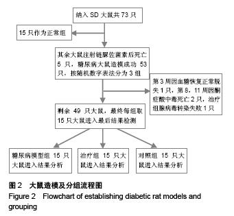
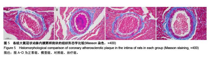
.jpg)
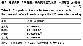
.jpg)