中国组织工程研究 ›› 2015, Vol. 19 ›› Issue (38): 6104-6108.doi: 10.3969/j.issn.2095-4344.2015.38.007

• 材料生物相容性 material biocompatibility • 上一篇    下一篇

羟基磷灰石表面修饰人工角膜钛支架的体内生物相容性

汪雪梅1,赵秋芳1,吴晓蓉2   

  1.  1陕西中医药大学附属医院眼科,陕西省咸阳市 712000; 2南昌大学第一附属医院眼科,江西省南昌市 330006)  
  • 通讯作者: 吴晓蓉,副主任医师,南昌大学第一附属医院眼科,江西省南昌市 330006
  • 作者简介:汪雪梅,女,1977年生,陕西省安康市人,2007年南昌大学毕业,硕士,主治医师,主要从事眼科疾病诊断与治疗研究。
  • 基金资助:

    国家自然科学基金(81360151):非天然构象H-AChR亚单位激活B细胞诱导眼肌型重症肌无力的机制研究

Hydroxyapatite surface modification of artificial cornea titanium scaffold: in vivo biocompatibility

Wang Xue-mei1, Zhao Qiu-fang1, Wu Xiao-rong2
  

  1. 1Department of Ophtalmology, Affiliated Hospital of Shaanxi University of Chinese Medicine, Xianyang 712000, Shaanxi Province, China;
     2Department of Ophtalmology, the First Affiliated Hospital of Nanchang University, Nanchang 330006, Jiangxi Province, China
  • Contact: Wu Xiao-rong, Associate chief physician, Department of Ophtalmology, the First Affiliated Hospital of Nanchang University, Nanchang 330006, Jiangxi Province, China
  • About author:Wang Xue-mei, Master, Attending physician, Department of Ophtalmology, Affiliated Hospital of Shaanxi University of Chinese Medicine, Xianyang 712000, Shaanxi Province, China
  • Supported by:

    the National Natural Science Foundation of China, No. 81360151

摘要:

背景:纯钛人工角膜支架在临床应用中的并发症发生率较高,因此寻找一种生物相容性高的人工角膜支架材料一直是国内外研究的重点和热点。

目的:观察羟基磷灰石表面修饰人工角膜钛支架的体内生物相容性。

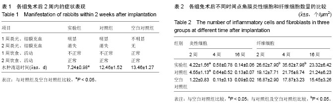
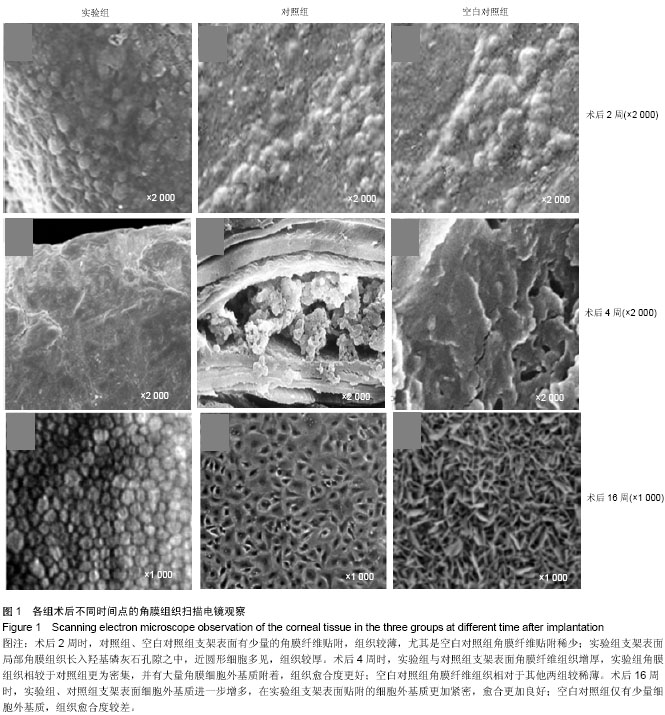
方法:取新西兰白兔27只,制作右眼角膜碱烧伤模型,造模后立即均分为3组,实验组右眼植入经过羟基磷灰石表面修饰的人工角膜钛支架,对照组右眼植入人工角膜钛支架,空白对照组右眼仅制备囊袋而不植入支架。术后2,4,16周取兔右眼角膜组织,进行病理组织学观察及扫描电镜观察。

结果与结论:术后16周,3组间炎性细胞与纤维细胞数量比较差异均无显著性意义。随着时间的延长,实验组角膜组织逐渐增多,纤维组织逐渐增厚,细胞外基质附着逐渐增加,角膜组织贴附密集度、细胞外基质附着密集度及组织愈合度均优于对照组及空白对照组。表明羟基磷灰石表面修饰人工角膜钛支架具有良好的生物相容性,可有效促进角膜细胞增生,有利于角膜血管化。

关键词: 生物材料, 材料相容性, 羟基磷灰石表面修饰法, 人工角膜, 钛支架, 生物相容性, 国家自然科学基金

Abstract:


BACKGROUND: Artificial cornea titanium scaffold has a higher complication rate in clinical usage; therefore, to seek a kind of artificial corneal scaffold with high biocompatibility is a hot spot.

OBJECTIVE: To explore the biocompatibility of the artificial cornea titanium scaffold with hydroxyapatite surface modification.

METHODS: Twenty-seven New Zealand white rabbits were enrolled to make alkali burn models of the right cornea and then randomized into three groups: in experimental group, artificial cornea titanium scaffold with hydroxyapatite surface modification was implanted into the right eye; in control group, artificial cornea titanium scaffold was implanted into the right eye; in blank group, no scaffold was implanted into the right eye. At 2, 4, 16 weeks after implantation, the right corneas were taken out for pathological observation and scanning electron microscope observation.

RESULTS AND CONCLUSION: At 16 weeks after implantation, there was no difference in the number of inflammatory cells and fibroblasts among the three groups. Over time, in the experimental group, the corneal tissues, fibrous tissues and adhered extracellular matrices were increased gradually. In addition, the experimental group had more adherent corneal tissues, extracellular matrices and better tissue healing than the control and blank groups. These findings indicate that the artificial cornea titanium scaffold with hydroxyapatite surface modification had good biocompatibility that can effectively promote proliferation of corneal cells and corneal vascularization.

中国组织工程研究杂志出版内容重点:生物材料;骨生物材料; 口腔生物材料; 纳米材料; 缓释材料; 材料相容性;组织工程

Key words: Durapatite, Cornea, Biocompatible Materials

中图分类号: