中国组织工程研究 ›› 2015, Vol. 19 ›› Issue (34): 5433-5437.doi: 10.3969/j.issn.2095-4344.2015.34.005

• 组织工程口腔材料 tissue-engineered oral materials • 上一篇    下一篇

崩瓷修补的临床试验及体外材料学拉伸强度实验

王 星1,齐 鲁2,顾振宇3,何惠宇1   

  1. 1新疆医科大学第一附属医院口腔修复科,新疆维吾尔自治区乌鲁木齐市 830054; 2新疆医科大学第二附属医院口腔科,新疆维吾尔自治区乌鲁木齐市 830063;3新疆兵团医院口腔科,新疆维吾尔自治区乌鲁木齐市 830011
  • 出版日期:2015-08-20 发布日期:2015-08-20
  • 作者简介:王星,女,1982年生,新疆维吾尔自治区呼图壁县人,汉族,2010年新疆医科大学毕业,硕士,主治医师,主要从事口腔修复学研究。

Clinical trial of collapsed repair and in vitro tensile strength test 

Wang Xing1, Qi Lu2, Gu Zhen-yu3, He Hui-yu1   

  1. 1Department of Prosthodontics, First Affiliated Hospital of Xinjiang Medical University, Urumqi 830054, Xinjiang Uygur Autonomous Region, China; Department of Stomatology, Second Affiliated Hospital of Xinjiang Medical University, Urumqi 830063, Xinjiang Uygur Autonomous Region, China; 3Department of Stomatology, Hospital of Xinjiang Production & Construction Corps, Urumqi 830011, Xinjiang Uygur Autonomous Region, China
  • Online:2015-08-20 Published:2015-08-20
  • About author:Wang Xing, Master, Attending physician, Department of Prosthodontics, First Affiliated Hospital of Xinjiang Medical University, Urumqi 830054, Xinjiang Uygur Autonomous Region, China

摘要:

背景:目前对瓷类修复体后崩瓷修补的研究较多,但缺乏系统性、横向的比较。

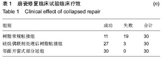
目的:通过临床试验及体外实验比较树脂常规粘接、硅烷偶联剂处理后树脂粘接及邻面开口式部分冠3种崩瓷修补方法的效果。

方法:①临床试验:将90例崩瓷患者随机均分为3组修补,分别采用树脂常规粘接、硅烷偶联剂处理后树脂粘接及邻面开口式部分冠3种崩瓷修补方法,对比3组1年后的修补成功率。②体外实验:将20个瓷试件均分为2组,分别进行树脂常规粘接及硅烷偶联剂处理后树脂粘接处理,检测两组剪切强度;将20个双层瓷试件均分为4组,其中3组黏结面分别进行喷砂、硅烷偶联剂、喷砂联合硅烷偶联剂处理,另1组不做任何处理(对照组),黏结后检测各组试件拉伸强度。

结果与结论:树脂常规粘接组、硅烷偶联剂处理后树脂粘接组及邻面开口式部分冠组修复成功率分别为37%,90%,100%。树脂常规粘接组与硅烷偶联剂处理后粘接组剪切强度分别为(13.978±0.343),(10.058±0.64) MPa,组间比较差异有显著性意义(P < 0.01)。对照组、喷砂组、硅烷偶联剂组、联合处理组的拉伸强度分别为(0.68±0.04),(1.00±0.02),(1.31±0.08),(1.09±0.04) kN,组间两两比较差异有显著性意义(P < 0.01)。表明硅烷偶联剂处理后树脂粘接及邻面开口式部分冠崩瓷修补效果优于树脂常规粘接。

中国组织工程研究杂志出版内容重点:组织构建;骨细胞;软骨细胞;细胞培养;成纤维细胞;血管内皮细胞;骨质疏松组织工程

关键词: 生物材料, 口腔生物材料, 崩瓷, 硅烷偶联剂, 邻面开窗式部分冠, 树脂

Abstract:

BACKGROUND: Currently, there are many studies on collapsed repair, but a systematic and horizontal comparison is not reported yet.

OBJECTIVE: To compare the effect of conventional resin adhesive, resin adhesive+silane coupling agent and adjacent surface open part of the crown on collapsed repair through clinical trial and in vitro experiment.

METHODS: (1) Clinical trial: 90 patients with porcelain collapse were randomized into three groups, and respectively treated with conventional resin adhesive, resin adhesive+silane coupling agent and adjacent surface open part of the crown. Success rate was measured and compared among three groups at 1 year after repair. (2) In vitro test: Twenty test specimens were equally divided into two groups, and treated with conventional resin adhesive and resin adhesive+silane coupling agent, respectively. Then, shear strength was detected in the two groups. Twenty double-crown specimens were equally divided into four groups. The first three groups were treated with sand blasting, silane coupling agent and their combination treatment, respectively; the rest group had no treatment (control group). After repair, the tensile strength of each specimen was detected.

RESULTS AND CONCLUSION: The success rate of collapsed repair was 37% for conventional resin adhesive, 90% for resin adhesive+silane coupling agent and 100% for adjacent surface open part of the crown. The shear strength was (13.978±0.343) MPa for the conventional resin adhesive and (10.058±0.64) MPa for resin adhesive+silane coupling agent, and there was a significant difference between two methods (P < 0.01). The tensile strength was (0.68±0.04) kN in the control group, (1.00±0.02) kN in the sand blasting group, (1.31±0.08) kN in silane coupling agent group, and (1.09±0.04) kN in the combination group, and there was a significant differences between groups (P < 0.01). Experimental results show that the silane coupling agent+resin adhesive treatment and adjacent surface open part of the crown are superior to conventional resin adhesive.

中国组织工程研究杂志出版内容重点:组织构建;骨细胞;软骨细胞;细胞培养;成纤维细胞;血管内皮细胞;骨质疏松组织工程

中图分类号: