中国组织工程研究 ›› 2021, Vol. 25 ›› Issue (10): 1489-1494.doi: 10.3969/j.issn.2095-4344.3052

• 组织工程骨材料Tissue-engineered bone • 上一篇    下一篇

骨形态发生蛋白9复合胶原基骨修复材料体外成骨性能与体内修复骨缺损

庄传记1,陈文昭2,江新民1   

  1. 1景德镇市第二人民医院骨科,江西省景德镇市 333000;2南昌大学第一附属医院骨科,江西省南昌市  330006
  • 收稿日期:2020-05-20 修回日期:2020-05-22 接受日期:2020-06-29 出版日期:2021-04-08 发布日期:2020-12-17
  • 通讯作者: 江新民,主任医师,景德镇市第二人民医院骨科,江西省景德镇市 333000
  • 作者简介:庄传记,男,1978年生,江西省鄱阳县人,硕士,副主任医师,主要从事关节、运动医学研究。
  • 基金资助:
    江西省健康委员会支持项目(20204356),课题名称:临床阳性体征与膝关节半月板损伤的相关性研究

Osteogenesis in vitro and bone defect repair in vivo of bone morphogenetic protein 9 composite collagen based bone repair material

Zhuang Chuanji1, Chen Wenzhao2, Jiang Xinmin1   

  1. 1Department of Orthopedics, Second People’s Hospital of Jingdezhen, Jingdezhen 333000, Jiangxi Province, China; 2Department of Orthopedics, the First Affiliated Hospital of Nanchang University, Nanchang 330006, Jiangxi Province, China
  • Received:2020-05-20 Revised:2020-05-22 Accepted:2020-06-29 Online:2021-04-08 Published:2020-12-17
  • Contact: Jiang Xinmin, Chief physician, Department of Orthopedics, Second People’s Hospital of Jingdezhen, Jingdezhen 333000, Jiangxi Province, China
  • About author:Zhuang Chuanji, Master, Associate chief physician, Department of Orthopedics, Second People’s Hospital of Jingdezhen, Jingdezhen 333000, Jiangxi Province, China
  • Supported by:
    the Project Supported by Jiangxi Health Committee, No. 20204356 (to ZCJ)

摘要:

文题释义:
骨形态发生蛋白:属于转化生长因子β超家族成员,因具有较强的诱导骨形成能力而得名,在人类中具有促成骨作用的主要有骨形态发生蛋白2、骨形态发生蛋白4、骨形态发生蛋白6与骨形态发生蛋白7,其中骨形态发生蛋白2与骨形态发生蛋白7目前已被临床用于促进脊柱融合。近些年的研究显示,骨形态发生蛋白成员中的骨形态发生蛋白9同样具有诱导骨形成与成骨分化的能力。
碱性磷酸酶:几乎存在于人体的任何组织,但主要集中于骨、肝脏、胆管、肾脏等,是成骨分化早期的重要标志性基因。

背景:胶原基骨修复材料具备良好的骨传导性、骨诱导性、降解性、机械性能与生物相容性,目前已被广泛应用于临床骨修复中,但其诱导成骨效果仍不及自体骨。
目的:将骨形态发生蛋白9(bone morphogenetic protein 9,BMP-9)复合于胶原基骨修复材料中,观察其诱导成骨的能力。
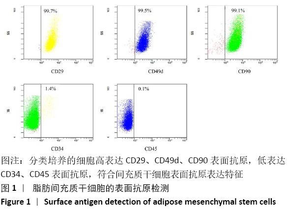
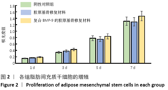
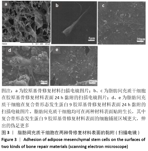
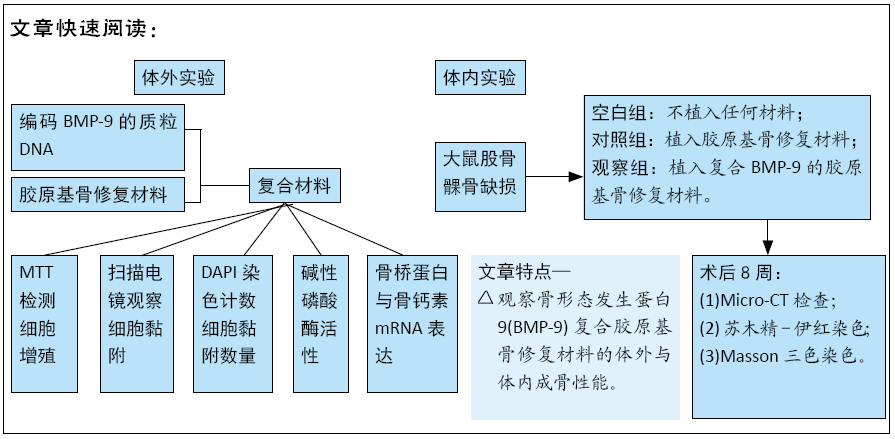
方法:将编码BMP-9的质粒DNA负载到胶原基骨修复材料中,构建复合BMP-9的胶原基骨修复材料。①体外实验:将SD大鼠脂肪间充质干细胞分别接种于复合BMP-9的胶原基骨修复材料与胶原基骨修复材料中,利用MTT法检测细胞增殖,扫描电镜观察细胞黏附,DAPI染色后计数细胞黏附数量,碱性磷酸酶活力试剂盒检测成骨诱导后细胞中的碱性磷酸酶活性,qRT-PCR检测成骨诱导后细胞中的骨桥蛋白与骨钙素mRNA表达量;②体内实验:在15只SD大鼠右后肢股骨内上髁制作直径3 mm、深3 mm的骨缺损,空白组不植入任何材料,对照组植入胶原基骨修复材料,观察组植入复合BMP-9的胶原基骨修复材料,术后8周取材,分别进行Micro-CT检查与组织学观察。实验方案经南昌大学实验动物科学中心伦理委员会批准。
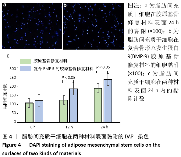
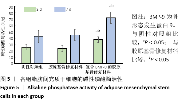
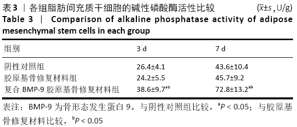
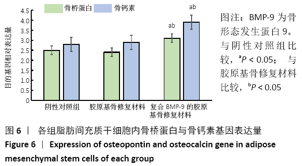
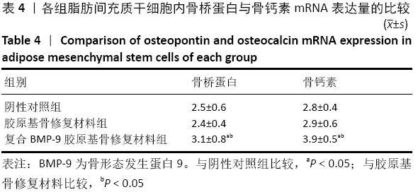
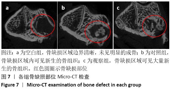
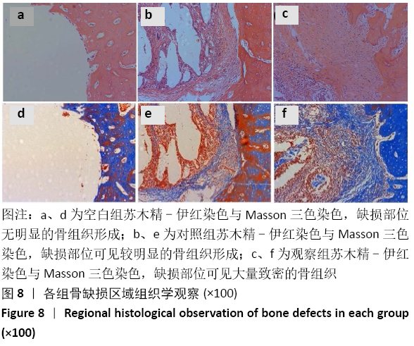
结果与结论:①体外实验显示,复合BMP-9胶原基骨修复材料中培养1,3,5,7 d的细胞增殖与胶原基骨修复材料比较差异无显著性意义(P﹥0.05);②体外共培养24 h后,在复合BMP-9胶原基骨修复材料表面黏附的细胞较多,并伸出较多的伪足,黏附细胞计数与铺展情况优于胶原基骨修复材料;③体外成骨诱导3,7 d后,复合BMP-9胶原基骨修复材料表面细胞的碱性磷酸酶活性高于胶原基骨修复材料(P < 0.05),成骨诱导7 d后的骨桥蛋白与骨钙素mRNA表达量高于胶原基骨修复材料(P < 0.05);④体内实验Micro-CT检查显示,观察组新骨生成量高于对照组、空白组(P < 0.05),对照组高于空白组(P < 0.05);⑤体内实验苏木精-伊红与Masson三色染色显示,观察组的骨修复效果优于对照组、空白组,对照组优于空白组;⑥结果表明,BMP-9可进一步提高胶原基骨修复材料的骨诱导性能。

https://orcid.org/0000-0002-4297-9304 (庄传记) 

中国组织工程研究杂志出版内容重点:生物材料;骨生物材料; 口腔生物材料; 纳米材料; 缓释材料; 材料相容性;组织工程

关键词: 骨, 材料, 胶原基骨, 骨形态发生蛋白, 骨缺损, 成骨, 骨诱导

Abstract: BACKGROUND: Collagen-based bone repair materials have been widely used in clinical bone repair due to their excellent osteoconductivity, osteoinduction, degradation, mechanical properties and biocompatibility, but their osteoinduction effect is not as good as that of autogenous bone.
OBJECTIVE: Bone morphogenetic protein 9 (BMP-9) was incorporated into collagen matrix to investigate its osteoinductive ability.
METHODS: The plasmid DNA encoding BMP-9 was loaded into collagen-based bone repair materials to construct a composite of collagen-based bone repair materials. (1) In vitro experiment: The Sprague-Dawley rat adipose mesenchymal stem cells were inoculated with BMP-9 composite collagen-based bone repair material and collagen-based bone repair material. Cell proliferation was detected by MTT assay, and the cell adhesion was observed by scanning electron microscope. Cell adhesion was counted after DAPI staining. Alkaline phosphatase activity was detected by alkaline phosphatase kit. Osteopontin and osteocalcin mRNA expression levels were detected by qRT-PCR. (2) In vivo experiment: Bone defects of 3 mm in diameter and 3 mm in depth were made in the medial epicondyle of the right hind limb of 15 Sprague-Dawley rats. No material was implanted in the blank group. Collagen-based bone repair material was implanted in the control group. BMP-9 composite collagen-based bone repair material was implanted in the observation group. Samples were obtained at 8 weeks after surgery. Micro-CT examination and histological observation were performed. This study was approved by the Ethics Committee of Laboratory Animal Science Center of Nanchang University. 
RESULTS AND CONCLUSION: (1) In vitro experiment showed that there was no significant difference between the proliferation of adipose tissue mesenchymal stem cell cultured for 1, 3, 5, and 7 days in collagen-based bone repair materials compounded with BMP-9 (P > 0.05). (2) After co-culture in vitro for 24 hours, more cells and more pseudopodia appeared on the surface of collagen-based bone repair material compounded with BMP-9. The number and spreading of adherent cells were better than that of collagen-based bone repair material. (3) After 3 and 7 days of osteogenic induction in vitro, the cell alkaline phosphatase activity of the collagen-based bone repair material compounded with BMP-9 was higher than that of the collagen-based bone repair material (P < 0.05). After 7 days of osteogenic induction, osteopontin and osteocalcin mRNA expression was higher than that of collagen-based bone repair material (P < 0.05). (4) In vivo micro-CT examination showed that the amount of new bone formation in the observation group was higher than that in the control group and the blank group    (P < 0.05), and that in the control group was higher than that in the blank group (P < 0.05). (5) In vivo hematoxylin-eosin staining and Masson staining showed that the bone repair effect of the observation group was better than that of the control and blank groups, and that in the control group was better than that in the blank group. (6) These findings confirmed that BMP-9 can further improve the bone induction properties of collagen-based bone repair materials. 

Key words: bone, material, collagen based bone, bone morphogenetic protein, bone defect, osteogenesis, osteoinduction

中图分类号: