中国组织工程研究 ›› 2015, Vol. 19 ›› Issue (49): 7920-7925.doi: 10.3969/j.issn.2095-4344.2015.49.009

• 脑及脊髓损伤动物模型 Animal models of brain and spinal cord injuries • 上一篇    下一篇

不同脊髓损伤模型大鼠行为学及形态学的变化

宋 伟1,2,段红梅3,饶家声3,赵 璨3,杨朝阳4   

  1. 1首都医科大学康复医学院,北京市 100068;2中国康复研究中心康复工程研究所,北京市 100068;3北京航空航天大学生物与医学工程学院,北京市 100191;4首都医科大学北京神经科学研究所,北京市 100054
  • 收稿日期:2015-09-25 出版日期:2015-11-30 发布日期:2015-11-30
  • 通讯作者: 杨朝阳,博士,副教授,硕士生导师,首都医科大学北京神经科学研究所,北京市 100054
  • 作者简介:宋伟,女,1963年生,北京市人,硕士,副教授,高级工程师,主要从事生物医学工程、康复工程研究。
  • 基金资助:

    国家科技支撑计划项目(2012BAI17B00)

Behavioral and morphological changes of different spinal cord injury rat models 

Song Wei1, 2, Duan Hong-mei3, Rao Jia-sheng3, Zhao Can3, Yang Zhao-yang4   

  1. 1Capital Medical University of Rehabilitation Medicine, Beijing 100068, China; 2Rehabilitation Engineering Research Institute of China Rehabilitation Research Center, Beijing 100068, China; 3Biological and Medical Engineering College of Beihang University, Beijing 100191, China; 4Beijing Institute of Neuroscience, Capital Medical University, Beijing 100054, China
  • Received:2015-09-25 Online:2015-11-30 Published:2015-11-30
  • Contact: Yang Zhao-yang, M.D., Associate professor, Master’s supervisor, Beijing Institute of Neuroscience, Capital Medical University, Beijing 100054, China
  • About author:Song Wei, Master, Associate professor, Senior engineer, Capital Medical University of Rehabilitation Medicine, Beijing 100068, China; Rehabilitation Engineering Research Institute of China Rehabilitation Research Center, Beijing 100068, China
  • Supported by:

    National Science and Technology Support Program, No. 2012BAI17B00

摘要:

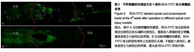
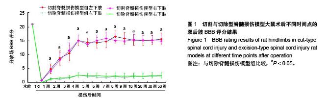
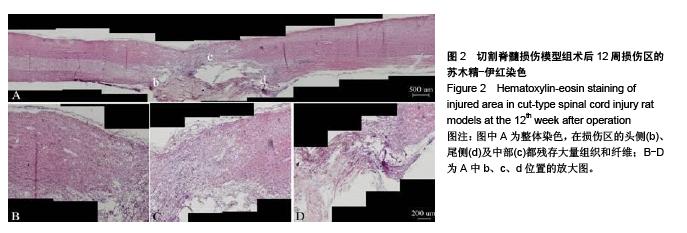
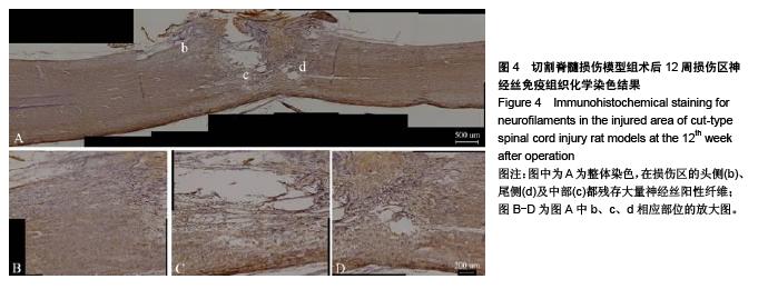
背景:在脊髓全断模型中有切割伤模型、切除伤模型,为了更好地选择脊髓损伤修复研究的全断动物模型,有必要对这两种模型进行比较及探讨。
目的:分析不同脊髓损伤模型的行为学及病理变化,选择适用于再生研究及治疗的脊髓损伤动物模型。
方法:将160只SD大鼠随机均分为2组,分别制作切割型脊髓损伤模型与切除型脊髓损伤模型,术后1,2,4,8,10,20,30,50周内进行后肢行为学BBB评分及组织化学、免疫组织化学染色,观察损伤区残余纤维情况;术后4,10,26,52周分别在大鼠脑皮质运动区与颈髓注射BDA-FITC示踪剂,观察损伤区残余纤维的连续性。
结果与结论:切割脊髓损伤模型组大鼠术后2-50周双后肢的BBB得分明显高于切除脊髓损伤模型组(P < 0.05);切割脊髓损伤模型组在损伤区残存大量组织和神经丝、胶质纤维酸性蛋白纤维,而切除脊髓损伤模型几乎没有残存纤维;不论是切割脊髓损伤模型还是切除脊髓损伤模型,皮质脊髓束均不能通过损伤区,终止于损伤区的头端;在切割脊髓损伤模型中,脊髓固有束重新进入尾侧端的宿主脊髓组织中,而在切除脊髓损伤模型中脊髓固有束终止于损伤区的头端。表明切除脊髓损伤模型更适用于脊髓损伤后再生效果及治疗手段或药物的筛选与评价。 


关键词: 实验动物模型, 脑及脊髓损伤动物模型, 脊髓损伤, 病理变化, 功能恢复, 神经示踪

Abstract:

BACKGROUND: Complete spinal cord transection models include cut-type injury and excision-type injury models. It is necessary to compare and discuss these two models, in order to select more appropriate animal models for studying spinal cord injury and repair.
OBJECTIVE: To analyze the behavioral and pathological changes of different spinal cord injury models and to select the animal models of spinal cord injury which are more suitable for studying regeneration and treatment of injured spinal cord.
METHODS: One hundred and sixty Sprague-Dawley rats were randomly divided into two groups to establish cut-type and excision-type spinal cord injury models. At 1, 2, 4, 8, 10, 20, 30 and 50 weeks after operation, rat hindlimb locomotor skills were evaluated using BBB scores; histochemical staining and immunohistochemical staining were conducted to observe the residual fibers in injured area; at 4, 10, 26 and 52 weeks after operation, BDA-FITC tracer agent was respectively injected into the motor area of the cerebral cortex and cervical cord of 
rats, to further observe the continuity of residual fibers in injured area.
RESULTS AND CONCLUSION: The BBB scores of rat hind limbs in cut-type spinal cord injury model group from 2 to 50 weeks after operation were significantly higher than those in excision-type spinal cord injury model group (P < 0.05). In cut-type spinal cord injury model group, there were a large amount of residual tissues, neurofilaments and glial fibrillary acidic protein-positive fibers in injured area, but there were almost no residual fibers in excision-type spinal cord injury model group. The corticospinal tract could pass through the injured area in neither cut-type spinal cord injury models nor excision-type spinal cord injury models and terminated at the cranial end of the injured area. In cut-type spinal cord injury models, the spinal cord propriospinal tract re-entered into the host spinal cord tissue at the caudal end, whereas the spinal cord propriospinal tract in excision-type spinal cord injury models terminated at the cranial end of the injured area. These results demonstrate that cut-type spinal cord injury models are more suitable for screening and evaluating the regeneration effect of treatments or drugs after spinal cord injury.  


中图分类号: