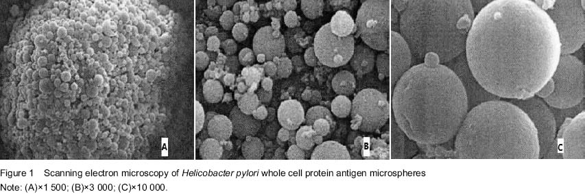
| [1] Aziz F, Sherwani SK, Akhtar SS, et al. Development of an in-house enzyme-linked immunosorbent assay based on surface whole cell antigen for diagnosis of Helicobacter pylori infection in patients with gastroduodenal ulcer disease. World J Microbiol Biotechnol. 2014;30(1): 305-315.
[2] Song W, Wang Y, Zhang L, et al. Preparation and evaluation of polysaccharide sulfates for inhibiting Helicobacter pylori adhesion. Carbohydr Polym. 2014;103:398-404.
[3] Liu YD, Li B, Gao WL, et al. Lumen of heavy moisture unit cell controlled release vaccine preparation and immune effect. Hebei Daxue Xuebao.2014;34(2):193-200.
[4] Gao Y, Wang X, Wan M, et al. Preparation of chitosan microspheres and its application in drug carrier. Func Mater. 2015;(2):2007-2012,2018.
[5] Zhang HQ, Gao QL, Guo HB, et al. Preparation of allogeneic bone load isoniazid-chitosan hydrogel microspheres and drug release characteristics in vitro and vivo. Zhongguo Zuzhi Goncgheng Yanjiu. 2012;16(3): 479-483.
[6] Li Qinhua, Wang Di. Preparation of calcium alginate and chitosan composite tissue engineering scaffolds with lyophilization. Zhongguo Zuzhi Goncgheng Yanjiu. 2012;16(29):5441-5444.
[7] Kucharska M, Walenko K, Lewandowska-Szumie? M, et al. Chitosan and composite microsphere-based scaffold for bone tissue engineering: evaluation of tricalcium phosphate content influence on physical and biological properties. J Mater Sci Mater Med. 2015;26(3):5464.
[8] Chen LY, Dang QZ, Liu CS, et al. Preparation and properties study of Chitosan-a solid dispersion loaded microspheres. Funct Mater. 2012;43(13):1762-1765, 1769.
[9] Prasad T, Shabeena EA, Vinod D, et al. Characterization and in vitro evaluation of electrospun chitosan/ polycaprolactone blend fibrous mat for skin tissue engineering. J Mater Sci Mater Med. 2015;26(1):5352.
[10] Liu W, Xie Y, Liu ZJ, et al. Releasing characteristics of Helicobacter pylori whole cell protein antigen chitosan microspheres in vitro. Zhongguo Zuzhi Goncgheng Yanjiu yu Linchuang Kangfu. 2011;15(38):7085-7089.
[11] Wu J, Ding SJ, Chen J, et al. Influence of attapulgite clay on releasing effect of Chitosan/sodium alginate microspheres. Huaxue Gongcheng Xueyuan Zazhi. 2014;(3):648-653.
[12] Hu YJ, Song L, Tian PC, et al. Preparation and releasing in vitro of Acyclovir ophthalmic chitosan gel in situ temperature-sensitive. Anhui Yiyao. 2014;18(1):30-32.
[13] Brachvogel RC, Hampel F, von Delius M. Self-assembly of dynamic orthoester cryptates. Nat Commun. 2015;6: 7129.
[14] Cui C, Qiao R, Zhang J. Monitoring residual platelet activity among patients with acute coronary syndrome post-pci by modified impedance whole blood platelet aggregation and release method. Clin Appl Thromb Hemost. 2015.
[15] Masuda Y, Asada K, Satoh R, et al. Capillin, a major constituent of Artemisia capillaris Thunb. flower essential oil, induces apoptosis through the mitochondrial pathway in human leukemia HL-60 cells. Phytomedicine. 2015;22(5): 545-552.
[16] Ujjwal RR, Purohit MP, Patnaik S, et al. General reagent free route to pH responsive polyacryloyl hydrazide capped metal nanogels for synergistic anticancer therapeutics. ACS Appl Mater Interfaces. 2015.
[17] Zur M, Cohen N, Agbaria R, et al. The biopharmaceutics of successful controlled release drug product: Segmental-dependent permeability of glipizide vs. metoprolol throughout the intestinal tract. Int J Pharm. 2015; 489(1-2):304-310.
[18] Jodar KS, Balcão VM, Chaud MV, et al. Development and Characterization of a Hydrogel Containing Silver Sulfadiazine for Antimicrobial Topical Applications. J Pharm Sci. 2015.
[19] Liu F, Sun J, Li YL, et al. Preparation and examination of BMP-2 loaded chitosan nanospheres in vitro. Shanghai Kou Qiang Yi Xue. 2015;24(2):147-150.
[20] Allam AN, Komeil IA, Fouda MA, et al. Preparation, characterization and in vivo evaluation of curcumin self-nano phospholipid dispersion as an approach to enhance oral bioavailability. Int J Pharm. 2015;489(1-2):117-123.
[21] Alcalá-Alcalá S, Benítez-Cardoza CG, Lima-Muñoz EJ, et al. Evaluation of a combined drug-delivery system for proteins assembled with polymeric nanoparticles and porous microspheres; characterization and protein integrity studies.Int J Pharm. 2015;489(1-2):139-147.
[22] Newton AM, Indana VL, Kumar J. Chronotherapeutic drug delivery of Tamarind gum, Chitosan and Okra gum controlled release colon targeted directly compressed Propranolol HCl matrix tablets and in-vitro evaluation. Int J Biol Macromol. 2015. |