| [1] 丁鹏,周彦恒,林久祥.自锁托槽矫治器的发展、分类及特点[J].口腔正畸学,2006,13(1):40-43.
[2] 贺钰,马俊青,赵红梅,等. Forsus和Dynamax矫治器对下颌骨生长改形的效果比较[J].广东牙病防治, 2010,(07)370-372.
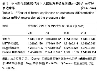
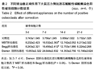
[3] 宋红,马秦, 陈永进.破骨细胞分化因子和破骨细胞生长抑制因子在牙周组织细胞中的表达[J].实用口腔医学杂志, 2009,25(5): 710-714.
[4] 张子扬,李玉如,黄统.拔除磨牙矫治安氏Ⅱ类错畸形患者的临床回顾性研究[J].国际口腔医学杂志, 2010,(02):141-145.
[5] 那宾,白雪芹,刘学恒. Damon-III矫治器治疗安氏Ⅱ类1分类错颌畸形患者硬组织变化的研究[J].国际口腔医学杂志, 2009, 36(3): 281-284.
[6] 朱红明,刘丹,章婷.拔除下颌磨牙矫治安氏Ⅲ类错关系的临床分析[J].口腔医学研究,2012, 28(4):370-372.
[7] 周欣荣. 自锁托槽矫治技术研究进展[J].北京口腔医学,2009,17(4):235-236.
[8] 牛白平.全新的正畸概念—Damon 系统:第一部分Damon系统的基本特点[J].实用口腔医学杂志,2008,24(4):608-611.
[9] Belibasakis GN, Bostanci N, Hashim A,et al. Regulation of ODF and OPG gene expression in human gingival fibroblasts and periodontal ligament cells by Porphyromonas gingivalis: A putative role of the Arg-gingipains. Microb Pathog. 2007;43(1): 46-53.
[10] Napimoga MH, Benatti BB, Lima FO, et al. Cannabidiol decreases bone resorption by inhibiting RANK/ODF expression and pro-inflammatory cytokines during experimental periodontitis in rats. Int Immunopharmacol. 2009;9(2):216-222.
[11] Irie K, Ekuni D, Yamamoto T, et al. A single application of hydrogen sulphide induces a transient osteoclast differentiation with ODF expression in the rat model. rchives of Oral Biology. 2009;54(8):723-729.
[12] Han G, Chen Y, Hou J, et al.Effects of simvastatin on relapse and remodeling of periodontal tissues after tooth movement in rats. Am J Orthod Dentofacial Orthop. 2010;138(5):550.e1-7;
[13] Ristic M, Vlahovic SM, Sasic M, et al. Clinical and microbiological effects of fixed orthodontic appliances on periodontal tissues in adolescents. Orthod Craniofacial Res,2007,10(4):187-195.
[14] 尹翰文,林娜,关键,等.双侧下颌支矢状劈开坚固内固定的三维有限元分析[J].上海口腔医学,2012, 21(2):194-198.
[15] Erkmen E, Simsek B, Yücel E, et al. Comparison of different fixation methods following sagittal split ramus osteotomies using three-dimensional finite elements analysis. Part 1: advancement surgery-posterior loading. Int J Oral Maxillofac Surg.2005;34(5):551-558.
[16] Reicheneder CA, Gedrange T, Lange A, et al. Editor's Summary, Q & A, Reviewer's Critique:Shear and tensile bond strength comparison of various contemporary orthodontic adhesive systems: An in-vitro study. Am J Orthod Dentofacial Orthop. 2009 ;135(4):422.e1-6.
[17] Chen SS, Greenlee GM, Kim JE, et al.Editor's Comment and Q&A: Systematic review of self-ligating brackets. Am J Orthod Dentofacial Orthop. 2010;137(6):726.e1-726.e18.
[18] Reddi D, Bostanci N, Hashim A, et al.Porphyromonas gingivalis regulates the ODF-OPG system in bone marrow stromal cells. Microbes and Infection. 2008;10(14-15): 1459-1468.
[19] Otsuka T, Kasai H, Yamaguchi K, et al.Enamel matrix derivative promotes osteoclast cell formation by ODF production in mouse marrow cultures. J Dent. 2005 Oct;33(9):749-755.
[20] Mogi M, Otogoto J. Expression of cathepsin-K in gingival crevicular fluid of patients with periodontitis.Archives of Oral Biology.2007;52(9)894-898.
[21] Jie Zhu, Chao Chen, Xue-nong Xing,et al.Effect of pioglitazone on expression of MMP-9 mRNA in human osteoclast-like cells induced by ODF and M-CSF.Bone.2010; 47(3):S418-S419.
[22] Wittrant Y, Theoleyre S, Couillaud S, et al. Regulation of osteoclast protease expression by RANKL. Biochem Biophys Res Commun. 2003;310(3):774-778.
[23] 王峰,林珠,李永明,等.机械力作用下人牙周膜细胞ODF及OCIF的表达及意义[J]. 实用口腔医学杂志 2005,21(01):85-87.
[24] Kawasaki K, Takahashi T, Yamaguchi M,et al.Effects of aging on ODF and OPG levels in gingival crevicular fluid during orthodontic tooth movemenL Orthod Craniofac Res.2006; 9(3):137-142. |