中国组织工程研究 ›› 2021, Vol. 25 ›› Issue (22): 3479-3486.doi: 10.3969/j.issn.2095-4344.3160

• 组织工程血管材料 tissue-engineered vascular materials • 上一篇    下一篇

构建预血管化细胞膜片及血管形成相关因子的表达

莫剑玲1,2,何少茹2,1,冯博文1,2,简敏桥2,张晓晖2,刘财盛2,梁一晶2,刘玉梅2,陈  亮2,周海榆2,刘艳辉2   

  1. 1南方医科大学第二临床医学院,广东省广州市   510515;2 广东省人民医院(广东省医学科学院),广东省广州市   510080
  • 收稿日期:2020-04-08 修回日期:2020-04-15 接受日期:2020-06-17 出版日期:2021-08-08 发布日期:2021-01-19
  • 通讯作者: 何少茹,博士,主任医师,博士生导师,广东省人民医院新生儿科(广东省医学科学院),广东省广州市 510080;南方医科大学第二临床医学院,广东省广州市 510515
  • 作者简介:莫剑玲,女,1993年生,广东省肇庆市人,汉族,南方医科大学在读硕士,主要从事干细胞与组织工程气管研究,以及儿童、新生儿呼吸疾病诊治。
  • 基金资助:
    国家自然科学基金项目(81671529),项目负责人:何少茹;广东省自然科学基金(2016A030313793),项目负责人:何少茹

Forming prevascularized cell sheets and the expression of angiogenesis-related factors

Mo Jianling1, 2, He Shaoru2, 1, Feng Bowen1, 2, Jian Minqiao2, Zhang Xiaohui2, Liu Caisheng2, Liang Yijing2, Liu Yumei2, Chen Liang2, #br# Zhou Haiyu2, Liu Yanhui2#br#   

  1. 1The Second School of Clinical Medicine, Southern Medical University, Guangzhou 510515, Guangdong Province, China; 2Guangdong Provincial People’s Hospital (Guangdong Academy of Medical Sciences), Guangzhou 510080, Guangdong Province, China
  • Received:2020-04-08 Revised:2020-04-15 Accepted:2020-06-17 Online:2021-08-08 Published:2021-01-19
  • Contact: He Shaoru, MD, Chief physician, Doctoral supervisor, Guangdong Provincial People’s Hospital (Guangdong Academy of Medical Sciences), Guangzhou 510080, Guangdong Province, China; The Second School of Clinical Medicine, Southern Medical University, Guangzhou 510515, Guangdong Province, China
  • About author:Mo Jianling, Master candidate, The Second School of Clinical Medicine, Southern Medical University, Guangzhou 510515, Guangdong Province, China; Guangdong Provincial People’s Hospital (Guangdong Academy of Medical Sciences), Guangzhou 510080, Guangdong Province, China
  • Supported by:
    Funding: the National Natural Science Foundation of China, No. 81671529 (to HSR); the Natural Science Foundation of Guangdong Province, No. 2016A030313793 (to HSR)

摘要:

文题释义:
细胞膜片技术:细胞膜片技术由日本学者OKANO等发明,该项技术具备许多优势,它不仅可以免去因使用胰蛋白酶消化给细胞带来伤害过程,保留了细胞外基质等结构,而且在维持细胞良好功能及提高细胞利用率中具有绝对优势。
组织工程:是应用细胞学和工程学的原理,研究开发用于修复或改善缺损组织、器官结构和功能的生物替代物的一门科学。组织工程技术是通过人体的基本单位——细胞,借助临时的三维支架和正常的生长方式建造出自然健康的自体组织,它的出现为组织缺损修复描绘了美好的前景。

背景:组织工程组织血管化问题难以解决,成为制约其临床应用的关键。
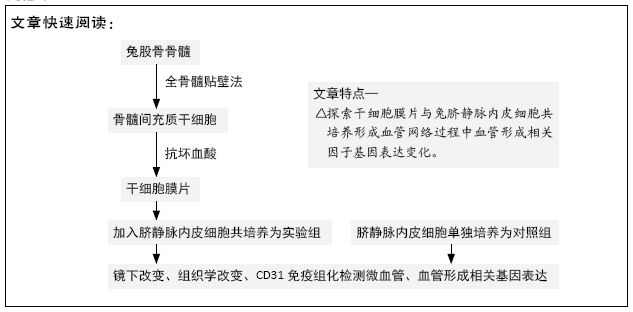
目的:探讨兔脐静脉内皮细胞与干细胞膜片共培养形成预血管化细胞膜片的可行性,分析其血管形成相关因子基因表达。
方法:采用全骨髓贴壁法体外分离培养兔骨髓间充质干细胞,经抗坏血酸作用形成干细胞膜片。实验组将干细胞膜片与兔脐静脉内皮细胞共培养以形成预血管化细胞膜片,以单独培养的兔脐静脉内皮细胞为对照,培养3,7,14 d时,光镜、苏木精-伊红染色观察细胞形态与组织学改变,RT-PCR检测血管形成相关因子mRNA表达,免疫组化染色观察血管化网络结构。
结果与结论:①光镜显示,实验组培养3 d后细胞发生迁移、重排,7 d后可见脐静脉内皮细胞之间发生“联系”,形成网状结构,14 d网状结构更加明显见;对照组细胞密度不断增加,形成细胞团,未见细胞发生重排、迁移形成网状结构;②苏木精-伊红染色显示,实验组培养3 d细胞排列不均匀,7 d时细胞呈条索状排列,14 d时细胞呈网状结构;对照组细胞呈铺路石样堆积且密度不断增大,无条索状、网状结构形成;③RT-PCR检测显示,实验组培养7 d的血管内皮生长因子mRNA表达高于对照组(P < 0.05),培养14 d的碱性成纤维细胞生长因子mRNA表达高于对照组(P < 0.05),培养7,14 d的血管生成素1 mRNA表达高于对照组(P < 0.05),培养3,7,14 d的血管生成素2 mRNA表达高于对照组(P < 0.05);④CD31免疫组化染色显示,实验组形成血管网状结构,对照组无网状结构形成;⑤结果表明,兔脐静脉内皮细胞与干细胞膜片共培养可以成功构建预血管化细胞膜片,显著表达血管形成相关基因。

关键词: 材料, 干细胞膜片, 骨髓间充质干细胞, 脐静脉内皮细胞, 抗坏血酸, 预血管化细胞膜片, 血管形成相关因子, 组织工程

Abstract: BACKGROUND: It is difficult to solve the problem of vascularization in tissue engineering, which has become the key to restrict its clinical application.
OBJECTIVE: To investigate the feasibility of forming a prevascularized cell sheet by co-culturing of rabbit umbilical vein endothelial cells and stem cell sheet, and to analyze the gene expression of angiogenesis related factors.
METHODS: Rabbit bone marrow mesenchymal stem cells were isolated and cultured in vitro, forming stem cell sheet by ascorbic acid. Prevascularized cell sheet was prepared by co-culture rabbit umbilical vein endothelial cells with stem cell sheet in the experimental group. Rabbit umbilical vein endothelial cells cultured separately were used as control group. At 3, 7, and 14 days, morphological and histological changes were observed by light microscopy and hematoxylin-eosin staining. mRNA expression of angiogenesis related factors was detected by RT-PCR, and the structure of vascularized network was observed by immunohistochemical staining.
RESULTS AND CONCLUSION: (1) The light microscopy showed that the cells of the experimental group migrated and rearranged at 3 days after culture. Rabbit umbilical vein endothelial cells "connected" and formed a network structure at 7 days. The network structure was more obvious at 14 days. The cell density of the control group increased continuously, forming a cell mass. No rearrangement or migration of cells was observed to form a network structure. (2) Hematoxylin-eosin staining showed that the cells in the experimental group were not uniformly arranged at 3 days, and the cells were arranged in cords at 7 days and reticulated at 14 days. The cells in the control group showed the accumulation of paving stones and the density increased continuously, without cords or reticular structures. (3) RT-PCR showed that mRNA expression of vascular endothelial growth factor was higher in the experimental group than in the control group at 7 days (P < 0.05). The mRNA expression of basic fibroblast growth factor was higher in the experimental group than that of the control group at 14 days (P < 0.05). The mRNA expression of Ang1 was higher in the experimental group than that of the control group at 7 and 14 days (P < 0.05). The mRNA expression of Ang2 was higher in the experimental group than that of the control group at 3, 7, and 14 days (P < 0.05). (4) Immunohistochemical staining of CD31 showed that the vascular network was formed in the experimental group, while no network was formed in the control group. (5) The results showed that rabbit umbilical vein endothelial cells co-cultured with stem cell membrane can successfully construct the pre-vascularized cell sheet, significantly expressing angiogenesis-related genes.

Key words: material, stem cell sheet, bone marrow mesenchymal stem cells, umbilical vein endothelial cells, ascorbic acid, prevascularized cell sheets, angiogenesis-related factors, tissue engineering

中图分类号: