中国组织工程研究 ›› 2021, Vol. 25 ›› Issue (2): 253-257.doi: 10.3969/j.issn.2095-4344.2953

• 组织构建实验造模 experimental modeling in tissue construction • 上一篇    下一篇

超短波对大鼠急性肺损伤炎症反应的影响

廖  源1,2,3,屈萌艰2,3,4,刘  静2,3,钟培瑞2,3,曾亚华2,3,王  婷2,3,肖  豪2,3,李  兰2,3,刘丹妮2,3,杨  璐2,3,周  君2,5,6    

  1. 1广州中医药大学针灸康复临床医学院,广东省广州市   510006;南华大学附属第一医院,2康复医学科,3康复医学实验室,湖南省衡阳市  421001;4美国克瑞顿大学,美国奥马哈  68178;5四川大学华西医院康复医学科,四川省成都市   610041;6康复医学四川省重点实验室,四川省成都市   610041
  • 收稿日期:2020-04-01 修回日期:2020-04-09 接受日期:2020-05-13 出版日期:2021-01-18 发布日期:2020-11-21
  • 通讯作者: 周君,博士,副教授,硕士生导师,南华大学附属第一医院,湖南省衡阳市 421001
  • 作者简介:廖源,女,1984年生,湖南省衡阳市人,汉族,广州中医药大学同等学力申请博士学位人员,主治医师,主要从事骨与关节康复、呼吸康复的临床与科研。
  • 基金资助:
    湖南省自然科学基金项目(2019JJ50544);衡阳市科技局项目(HKF201947209);南华大学附属第一医院亚专科重点项目(2017ZD007)

Effects of ultrashort wave on inflammatory response in rats with acute lung injury

Liao Yuan1, 2, 3, Qu Mengjian2, 3, 4, Liu Jing2, 3, Zhong Peirui2, 3, Zeng Yahua2, 3, Wang Ting2, 3, Xiao Hao2, 3, Li Lan2, 3, Liu Danni2, 3, Yang Lu2, 3, Zhou Jun2, 5, 6   

  1. 1Acupuncture and Rehabilitation College of Guangzhou University of Chinese Medicine, Guangdong Province, China; 2Department of Rehabilitation Medicine, 3Laboratory of Rehabilitation Medicine, First Affiliated Hospital of Nanhua University, Hunan Province, China; 4Creighton University, Omaha, NE 68178, USA; 5Department of Rehabilitation Medicine, West China Hospital, Sichuan University, Sichuan Province, China; 6Sichuan Provincial Key Laboratory of Rehabilitation Medicine, Sichuan Province, China
  • Received:2020-04-01 Revised:2020-04-09 Accepted:2020-05-13 Online:2021-01-18 Published:2020-11-21
  • Contact: Zhou Jun, MD, Associate professor, Master’s supervisor, Department of Rehabilitation Medicine, First Affiliated Hospital of Nanhua University, Hengyang 421001, Hunan Province, China
  • About author:Liao Yuan, Attending physician, Acupuncture and Rehabilitation College of Guangzhou University of Chinese Medicine, Guangzhou 510006, Guangdong Province, China
  • Supported by:
    the Natural Science Foundation of Hunan Province, No. 2019JJ50544 ; the Project of Hengyang Science and Technology Bureau, No. HKF201947209 ; Key Sub-Specialty Project of the First Affiliated Hospital of South China University, No. 2017ZD007

摘要:

文题释义:
急性肺损伤:心源性以外的各种肺内、外致病因素导致的急性、进行性、缺氧性呼吸衰竭。急性肺损伤主要为肺泡上皮和毛细血管内皮细胞损伤,表现为弥漫性肺间质及肺泡水肿,发病时可导致急性呼吸功能障碍。急性肺损伤最严重的情况就是急性呼吸窘迫综合征。
超短波治疗:是一种临床常用的物理因子治疗,具有扩张血管、使血管壁通透性增高、改善血液循环、促进炎症吸收、减轻水肿、增强组织营养及加强局部组织代谢的生理效应。超短波常用于治疗感染性疾病,有一定的抗炎作用。

背景:超短波常用于肺部感染的对症治疗,有一定抗炎作用。炎症反应失控是急性肺损伤的重要发病机制,抑制炎症反应是控制急性肺损伤的重要策略。
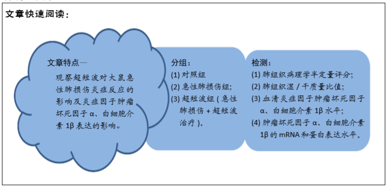
目的:观察超短波对大鼠急性肺损伤炎症反应的影响及炎症因子肿瘤坏死因子α、白细胞介素1β表达的影响。
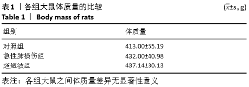
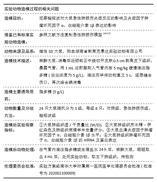
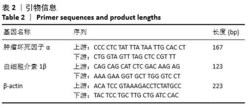
方法:24 只3月龄雄性SD大鼠随机分为对照组、急性肺损伤组、超短波组,每组8 只。急性肺损伤组与超短波组大鼠通过气管内滴注脂多糖复制急性肺损伤模型,对照组大鼠气管内滴注等量生理盐水。超短波组在大鼠滴注脂多糖即刻及滴注后4,8 h给予超短波干预,每次 15 min。24 h后取材,对大鼠肺组织进行苏木精-伊红染色,肺组织病理学半定量评分和肺组织湿/干质量比值比较;ELISA检测血清炎症因子肿瘤坏死因子α、白细胞介素1β水平,RT-PCR、Western-blot 分别检测肿瘤坏死因子α、白细胞介素1β的mRNA和蛋白表达水平。实验方案经南华大学附属第一医院医学伦理委员会批准(批准号为202002100009)。
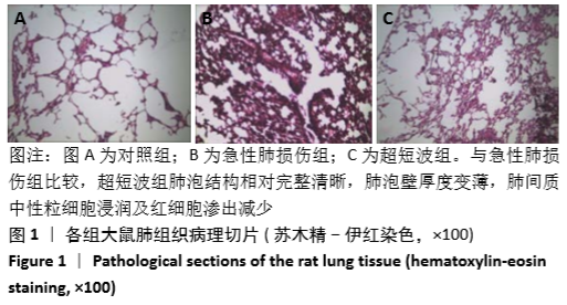
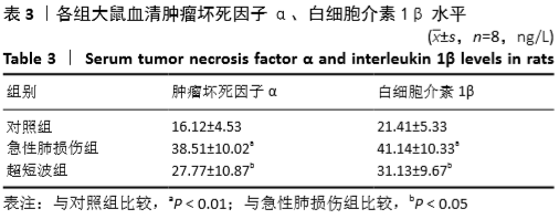
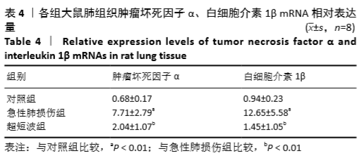
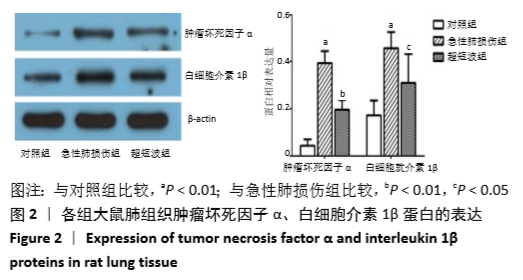
结果与结论:①肺组织湿/干质量比:急性肺损伤组较对照组明显升高(P < 0.01),超短波组比急性肺损伤组降低,但差异无显著性意义;②病理表现:急性肺损伤组大鼠肺组织结构破坏明显、肺泡隔大小不一,肺泡壁不完整、变厚断裂,肺间质可见大量中性粒细胞浸润和部分红细胞渗出;超短波组肺泡结构相对完整清晰,肺间质炎症细胞浸润及红细胞渗出减少;③肺组织病理学半定量评分:急性肺损伤组较对照组明显升高(P < 0.01);超短波组较急性肺损伤组明显降低(P < 0.01);④急性肺损伤组的血清肿瘤坏死因子α、白细胞介素1β较对照组明显升高(均P < 0.01),超短波组较急性肺损伤组明显降低(均P < 0.05);⑤超短波组肺组织肿瘤坏死因子α、白细胞介素1β mRNA和蛋白较急性肺损伤组明显降低(均P < 0.01或P < 0.05);⑥结果说明,超短波可能通过下调炎症因子肿瘤坏死因子α、白细胞介素1β的表达,抑制急性肺损伤大鼠肺组织的炎症反应。
https://orcid.org/0000-0002-1580-5747 (廖源) 

中国组织工程研究杂志出版内容重点:组织构建;骨细胞;软骨细胞;细胞培养;成纤维细胞;血管内皮细胞;骨质疏松;组织工程

关键词: 急性肺损伤, 超短波, 炎症反应, 因子, 模型, 鼠, 实验

Abstract: BACKGROUND:  Ultrashort wave can inhibit the inflammatory response and is often used in symptomatic treatment of pulmonary infection. Uncontrolled inflammatory response is an important pathogenesis of acute lung injury. Inhibiting inflammation is an important strategy for controlling acute lung injury.
OBJECTIVE: To observe the effects of ultrashort wave on inflammatory response and the expression of tumor necrosis factor-α (TNF-α) and interleukin-1β (IL-1β) in rats with acute lung injury.
METHODS: Twenty-four 3-month-old male Sprague-Dawley rats were randomly divided into control group, acute lung injury group (model group) and ultrashort wave group (n=8 per group). Rats in the acute lung injury group and the ultrashort wave group were given intratracheal lipopolysaccharide to replicate the acute lung injury model. Rats in the control group were given intratracheal infusion of the same amount of normal saline. In the ultrashort wave group, rats were given ultrashort wave intervention immediately, 4 and 8 hours after lipopolysaccharide infusion, once for 15 minutes. Experimental animals were sacrificed 24 hours after intratracheal infusion of lipopolysaccharide or normal saline. The lung tissues of rats were compared by hematoxylin-eosin staining, lung histopathological semi-quantitative score and lung wet/dry weight ratio (W/D). Serum levels of inflammatory factors TNF-α and IL-1β were detected by ELISA, and mRNA and protein expressions of TNF-α and IL-1β were detected by RT-PCR and western blot, respectively. The study protocol was approved by the First Affiliated Hospital of Nanhua University, approved No.202002100009.
RESULTS AND CONCLUSION: Lung W/D ratio in the acute lung injury group was significantly higher than that in the control group (P < 0.01), whereas the ratio in the ultrashort wave group was lower than that in the acute lung injury group, but there was no significant difference between the two groups. Pathological manifestations: In the model group, the lung tissue structure was obviously damaged, with different sizes of alveolar septa, the alveolar wall was incomplete, thickened and fractured, a large number of neutrophils were seen in the lung interstitium, and some red blood cells were exudated. In the ultrashort wave group, compared with the model group, the alveolar structure was relatively complete and clear, and the infiltration of inflammatory cells and red blood cell exudation from the lung interstitium were reduced. Semi-quantitative histopathological score of the lung was significantly higher in the model group than the control group (P < 0.01), but significantly reduced in the ultrashort wave group compared with the model group (P < 0.01). Serum TNF-α and IL-1β levels in the model group were significantly higher than those in the control group (both P < 0.01). After ultrashort wave exposure, the levels of serum TNF-α and IL-1β significantly decreased compared to the model group (both P < 0.05).  After ultrashort wave exposure, the mRNA and protein expressions of TNF-α and IL-1β significantly decreased compared to the model group (P < 0.01 or P < 0.05). To conclude, ultrashort wave may inhibit the inflammatory response of the lung tissue in rats with acute lung injury by down-regulating the expression of inflammatory cytokines, TNF-α and IL-1β. 

Key words: acute lung injury, ultrashort wave, inflammatory response, factor, model, rat, experiment

中图分类号: