中国组织工程研究 ›› 2020, Vol. 24 ›› Issue (35): 5595-5599.doi: 10.3969/j.issn.2095-4344.2882

• 骨组织构建 bone tissue construction • 上一篇    下一篇

富血小板血浆促进胫骨骨折模型兔的骨愈合

任  军,赵  岩,肖  彬,马  超,王新苛,郝亚斌,成  杰   

  1. 新疆医科大学第一附属医院昌吉分院骨科,新疆维吾尔自治区昌吉市  831100

  • 收稿日期:2019-11-08 修回日期:2019-11-29 接受日期:2020-01-10 出版日期:2020-12-18 发布日期:2020-10-16
  • 通讯作者: 赵岩,博士,主任医师,新疆医科大学第一附属医院昌吉分院骨科,新疆维吾尔自治区昌吉市 831100
  • 作者简介:任军,男,1973年生,新疆维吾尔自治区奇台县人,汉族,2007年新疆医科大学毕业,副主任医师,主要从事创伤运动医学及脊柱微创研究。
  • 基金资助:
    昌吉市科学研究与技术开发计划项目

Platelet-rich plasma promotes the healing of tibial fracture in rabbits

Ren Jun, Zhao Yan, Xiao Bin, Ma Chao, Wang Xinke, Hao Yabin, Cheng Jie   

  1. Department of Orthopedics, Changji Branch, the First Affiliated Hospital of Xinjiang Medical University, Changji 831100, Xinjiang Uygur Autonomous Region, China

  • Received:2019-11-08 Revised:2019-11-29 Accepted:2020-01-10 Online:2020-12-18 Published:2020-10-16
  • Contact: Zhao Yan, MD, Chief physician, Department of Orthopedics, Changji Branch, the First Affiliated Hospital of Xinjiang Medical University, Changji 831100, Xinjiang Uygur Autonomous Region, China
  • About author:Ren Jun, Associate chief physician, Department of Orthopedics, Changji Branch, the First Affiliated Hospital of Xinjiang Medical University, Changji 831100, Xinjiang Uygur Autonomous Region, China
  • Supported by:
    the Science and Technology Development Plan of Changji

摘要:

文题释义:

富血小板血浆:通过高速离心的方法从全血中提取出来的血小板浓缩液,含有高浓度血小板和自身生长因子的血浆。

骨折愈合:是骨折断端之间组织修复的连续过程,成骨细胞与破骨细胞,联合多种细胞和生长因子参与的一系列复杂的生化反应,各种生物活性因子之间可以通过磷酸化等变化联系在一起。

骨折塑形期为了符合人体生理结构要求,具有更牢固的结构和良好的功能,骨性骨痂通过成骨细胞、破骨细胞的协调作用下成为成熟的板层骨,使骨折两断端再次完全连接起来,髓腔再通,骨折恢复到与原骨组织一样的结构,达到完全愈合。   

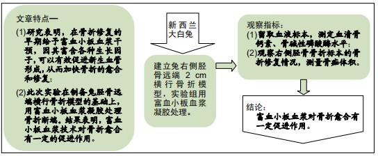
背景:骨折具有自我愈合周期长、延迟愈合等问题。生物组织具有自我修复的潜能,那么有没有办法将组织自身修复能力调动起来,为生物体自身修复所用呢?富血小板血浆技术正是解决这一难题的有效方式之一。

目的:探讨自体富血小板血浆对兔胫骨骨折愈合的促进作用。
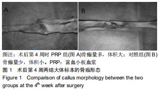
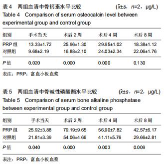
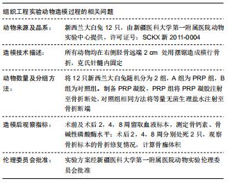
方法选取新西兰大白兔12只,随机分为富血小板血浆组及对照组,每组6只。所有动物均在右侧胫骨远端2 cm处用摆锯造成横行骨折,克氏针髓内固定。富血小板血浆组将制备的富血小板血浆凝胶注射至骨折断处,对照组相同方法将等量无菌生理盐水注射至骨折断端。术前及术后248周留取血液标本,测定骨钙素、骨碱性磷酸酶水平;术后248周分别处死2只,观察骨折标本的骨折修复情况,计算骨痂体积。

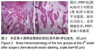
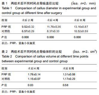
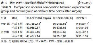
结果与结论:①术后各时点,富血小板血浆组骨痂直径均大于对照组(P < 0.05);②术后第4周,富血小板血浆组骨痂体积明显大于对照组(P < 0.05);术后第8周时,富血小板血浆组骨痂体积大于对照组,但差异无显著性意义(P > 0.05);③术后第4周富血小板血浆组骨小梁平均宽度、骨小梁面积比及血管数目均优于对照组(P < 0.05);术后第8周2组数据差异无显著性意义(P > 0.05);④2组血清中骨钙素水平在术后第4周时达到最高,可维持高浓度一段时间,富血小板血浆组最高值大于对照组(P < 0.05);⑤富血小板血浆组中血清碱性磷酸酶水平始终高于对照组,说明富血小板血浆在骨折愈合过程中对碱性磷酸酶的影响始终存在;⑥提示富血小板血浆技术对骨折愈合有一定的促进作用,可为骨科临床骨折不愈合或延迟愈合的治疗提供新思路、新方法。

ORCID: 0000-0002-8527-6895(任军)

中国组织工程研究杂志出版内容重点:组织构建;骨细胞;软骨细胞;细胞培养;成纤维细胞;血管内皮细胞;骨质疏松;组织工程

关键词: 骨, 骨折, 富血小板血浆, 兔, 动物, 内固定, 骨痂

Abstract:

BACKGROUND: There are many questions about fracture healing, such as long self-healing cycles and delayed healing. Biological tissue has the potential for self-repair, so is there a way to mobilize the tissue’s self-repair ability for the organism’s self-repair? Platelet-rich plasma technology is one of the effective ways to solve this problem.

OBJECTIVE: To study the effect of autologous platelet-rich plasmaon the healing of tibial fracture in rabbits.

METHODS: Twelve New Zealand white rabbits were randomly divided into experimental group and control group, with six rabbits in each group. All animals were transversely fractured with a pendulum saw at a site of 2 cm distal to the right tibia, followed by fixation with Kirschner wire. In the experimental group, the platelet-rich plasma gel was injected into the fracture site, and in the control group the same amount of sterile saline was injected into the fracture site. Blood samples from rabbit models were taken before and at 2, 4, and 8 weeks after operation. The contents of osteocalcin and bone alkaline phosphatase were measured. Two rats from each group were killed at 2, 4, and 8 weeks after operation. Repair of fractures and volume of callus were observed and calculated, respectively.

RESULTS AND CONCLUSION: The diameter of the callus in the experimental group was significantly larger than that of the control group at each observation time after operation (P < 0.05). Compared with the control group, the volume of the callus was significantly larger in the experimental group at 4 weeks postoperatively (P < 0.05), and still larger but not significantly at 8 weeks postoperatively (P > 0.05). The average width of trabecular bone, area ratio of trabecular bone and amount of blood vessels in the experimental group were better than those in the control group at 4 weeks postoperatively (P < 0.05); however, there was no significant difference between the two groups at 8 weeks postoperatively (P > 0.05). Serum osteocalcin in the two groups reached the highest level at 4 weeks postoperatively, and could keep a high level for a period of time. The peak level of serum osteocalcin in the experimental group was significantly higher than that in the control group (P < 0.05). The serum alkaline phosphatase level in the experimental group was always higher than that in the control group, indicating that platelet-rich plasma always has an effect on alkaline phosphatase level during fracture healing. To conclude, platelet-rich plasma has a certain promoting effect on the healing of tibial fracture, providing new ideas for treating fracture nonunion or delayed healing.

Key words: bone, fracture, platelet-rich plasma, rabbit, animal, internal fixation, callus

中图分类号: