中国组织工程研究 ›› 2020, Vol. 24 ›› Issue (21): 3299-3303.doi: 10.3969/j.issn.2095-4344.2678

• 骨与关节生物力学Bone and joint biomechanics • 上一篇    下一篇

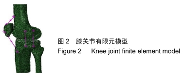
步态周期下半月板损伤对膝关节生物力学性能的影响

吴  铮,任  静,万建杉,孙  嵘,武成聪,刘克廷,刘  涛,欧  华   

  1. 曲靖市第一人民医院骨一科,云南省曲靖市  655000
  • 收稿日期:2019-11-15 修回日期:2019-11-19 接受日期:2019-12-13 出版日期:2020-07-28 发布日期:2020-04-14
  • 作者简介:吴铮,男,1980年生,云南省曲靖市人,在读硕士,副主任医师,主要从事骨科方面的研究。
  • 基金资助:
    云南省教育厅科学研究基金项目(2016ZDX074)

Effect of meniscus injury on the biomechanical properties of knee joint under gait cycle

Wu Zheng, Ren Jing, Wan Jianshan, Sun Rong, Wu Chengcong, Liu Keting, Liu Tao, Ou Hua   

  1. First Department of Orthopedics, First People’s Hospital of Qujing, Qujing 655000, Yunnan Province, China
  • Received:2019-11-15 Revised:2019-11-19 Accepted:2019-12-13 Online:2020-07-28 Published:2020-04-14
  • About author:Wu Zheng, Master candidate, Associate chief physician, First Department of Orthopedics, First People’s Hospital of Qujing, Qujing 655000, Yunnan Province, China
  • Supported by:
    the Science Research Foundation of Education Department of Yunnan Province, No. 2016ZDX074

摘要:

文题释义:

半月板撕裂:膝关节内半月型纤维软骨破裂,撕裂原因主要是由于膝半屈或全屈位下的扭转力所造成。半月板分为内侧半月板和外侧半月板,内侧半月板较大且固定,外侧半月板较小,实验主要研究外侧半月板撕裂对力学机制的影响。

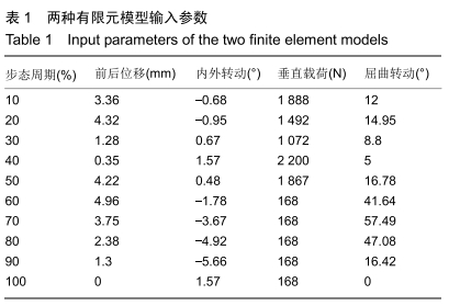
动态有限元分析:将人体正常完整步态周期作为边界条件施加在膝关节半月板模型中,观察在完整步态周期下半月板以及胫骨软骨的应力变化趋势及所受应力值大小。

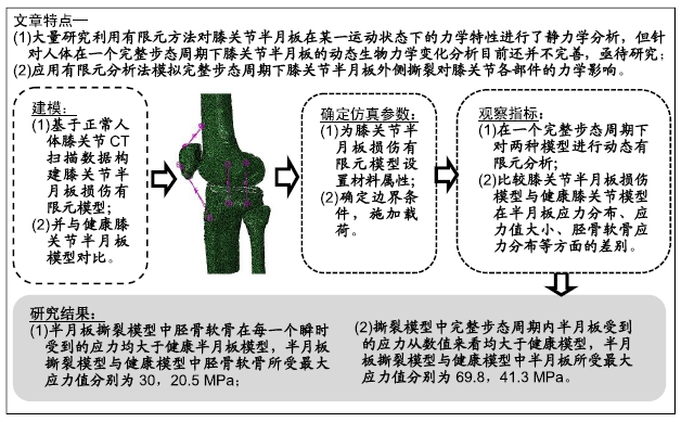
背景:目前国内外对膝关节半月板的生物力学分析十分广泛,但大多集中于对膝关节屈曲运动状态下的研究,针对完整步态周期下膝关节半月板生物力学的有限元分析还不完善。

目的:通过对比外侧半月板撕裂模型与健康半月板模型,了解完整步态周期下半月板损伤后的生物力学变化机制。

方法:以健康成年人膝关节CT扫描数据为基础,建立包括胫-股骨、半月板、关节软骨在内的健康膝关节有限元模型,并在健康模型基础上进一步构建膝关节外侧半月板撕裂模型,探究在完整步态周期下膝关节外侧半月板撕裂的生物力学机制,并与健康膝模型进行对比。

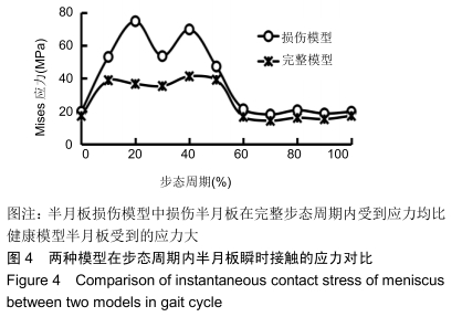
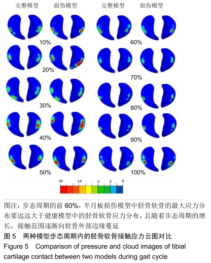
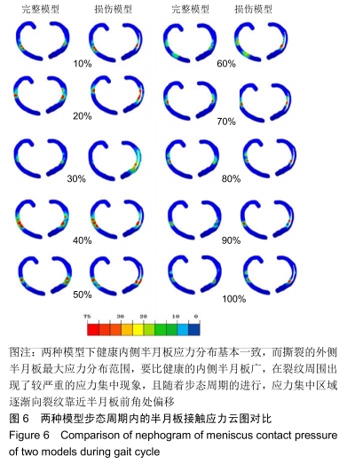
结果与结论:①两种模型完整步态周期内的胫骨软骨瞬时应力变化趋势一致,但半月板撕裂模型中胫骨软骨在每一个瞬时受到的应力值均大于健康半月板模型,半月板撕裂模型与健康模型中胫骨软骨所受最大应力值分别为30,20.5 MPa;②两种模型完整步态周期内的半月板瞬时应力变化趋势是一致的,但撕裂模型中完整步态周期内半月板受到的应力均大于健康模型,半月板撕裂模型与健康模型中半月板所受最大应力值分别为69.8,41.3 MPa;③在步态周期的前60%,半月板撕裂模型中的胫骨软骨最大应力分布远大于健康模型,且随着步态周期的增长,接触范围逐渐向软骨外部边缘蔓延;在步态周期的60%以后,作用在胫骨软骨上的应力较小,最大应力的分布范围也比较小;④两种模型中健康内侧半月板应力分布基本一致,而撕裂的外侧半月板最大应力分布范围较健康内侧半月板广,在裂纹周围出现了较严重应力集中现象,且随着步态周期的进行,应力集中区域逐渐向裂纹靠近半月板前角处偏移;⑤结果表明半月板是人体膝关节中重要的承重部件,从生物力学角度可以较为直观地观察到半月板损伤对人体膝关节的危害。

ORCID: 0000-0002-2155-0058(吴铮)

中国组织工程研究杂志出版内容重点:人工关节;骨植入物;脊柱骨折;内固定;数字化骨科;组织工程

关键词: 半月板, 生物力学, 应力, 有限元分析, 应力分布, 胫骨软骨, 完整步态周期, 数字化骨科

Abstract:

BACKGROUND: The biomechanical characteristics of knee meniscus have been studied extensively at home and abroad, but most of them focus on the knee flexion motion. The finite element analysis of biomechanics of knee joint meniscus under the complete gait cycle is not yet perfect.

OBJECTIVE: To understand the mechanism of biomechanical changes after meniscus injury in the complete gait cycle by comparing the lateral meniscus tear model with the healthy meniscus model.

METHODS: Based on the CT scan data of healthy adult knee joints, a finite element model of healthy knee joint including tibia, meniscus and articular cartilage was established. The lateral meniscus tear of knee joint was constructed based on the healthy model. The biomechanical mechanism of lateral meniscus tear in the knee during complete gait cycle was explored and compared with the healthy knee model.

RESULTS AND CONCLUSION: (1) The instantaneous stress variation of the tibia cartilage during the complete gait cycle was consistent in both models. The tibial cartilage stress at each instant in the meniscus tear model was higher than that of the healthy meniscus model. The maximum stress values of tibia cartilage in the meniscus tear model and the healthy meniscus model was 30 and 20.5 MPa. (2) The instantaneous stress variation of the meniscus during the complete gait cycle was consistent in both models. The meniscus stress at each instant in the meniscus tear model was higher than that of the healthy meniscus model. The maximum stress values of meniscus in the meniscus tear model and the healthy meniscus model was 69.8 and 41.3 MPa. (3) In the first 60% of the gait cycle, the maximum stress distribution of the tibia cartilage in the meniscus tear model was much larger than that in the healthy model, and as the gait cycle grew, the contact range gradually spread to the outer edge of the cartilage. After 60% of the gait cycle, the stress on the tibia cartilage was small, and the distribution range of the maximum stress was also small. (4) The stress distribution of the healthy medial meniscus was basically the same in the two models, while the maximum stress distribution of the torn outer meniscus was wider than that of the healthy medial meniscus. A more severe stress concentration phenomenon occurred around the crack, and with the gait cycle, the stress concentration area gradually shifted toward the crack near the anterior corner of the meniscus. (5) These results suggest that the meniscus is an important load-bearing component in human knee joint. From the perspective of biomechanics, the hazard of the meniscus injury on the human knee joint can be observed more intuitively.

Key words: meniscus, biomechanics, stress, finite element analysis, stress distribution, tibia cartilage, whole gait cycle, digital orthopedics

中图分类号: