中国组织工程研究 ›› 2020, Vol. 24 ›› Issue (9): 1400-1404.doi: 10.3969/j.issn.2095-4344.2512

• 骨与关节图像与影像 bone and joint imaging • 上一篇    下一篇

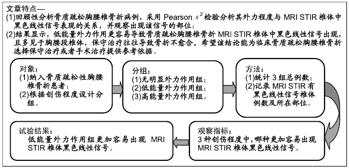
骨质疏松性胸腰椎骨折MRI STIR黑色线性信号与外力程度的关系

钟远鸣1,罗  满2,唐福波1,唐  成3   

  1. 1广西中医药大学第一附属医院,广西壮族自治区南宁市  530001;2广西国际壮医医院,广西壮族自治区南宁市  530001;3柳州市中医医院,广西壮族自治区柳州市  545001
  • 收稿日期:2017-11-20 修回日期:2017-11-24 接受日期:2019-02-06 出版日期:2020-03-28 发布日期:2020-02-12
  • 作者简介:钟远鸣,男,1965年生,广西壮族自治区南宁市人,壮族,广西中医药大学毕业,硕士,博士生导师,主任医师,主要从事脊柱脊髓相关疾病诊治研究。
  • 基金资助:


Relationship between a linear black signal area of STIR image in MRI of osteoporotic thoracolumbar fracture and the size of external force

Zhong Yuanming1, Luo Man2, Tang Fubo1, Tang Cheng3   

  1. 1The First Affiliated Hospital of Guangxi University of Chinese Medicine, Nanning 530001, Guangxi Zhuang Autonomous Region, China; 2Guangxi International Zhuang Medical Hospital, Nanning 530001, Guangxi Zhuang Autonomous Region, China; 3Liuzhou Traditional Chinese Medicine Hospital, Liuzhou 545001, Guangxi Zhuang Autonomous Region, China
  • Received:2017-11-20 Revised:2017-11-24 Accepted:2019-02-06 Online:2020-03-28 Published:2020-02-12
  • About author:Zhong Yuanming, Master, Doctoral supervisor, Chief physician, The First Affiliated Hospital of Guangxi University of Chinese Medicine, Nanning 530001, Guangxi Zhuang Autonomous Region, China

摘要:

文题释义:
MRI STIR椎体内黑色线性信号:指的是在MRI T2压脂像中,骨折椎体内会出现高信号,在骨折椎体高信号中有线性低信号出现,且在椎体内,为椎体压缩性骨折的特殊表现,出现骨折椎体内黑色线性信号往往提示着骨折保守治疗不愈合。

背景:随着社会老龄化加重,骨质疏松椎体骨折患者日益增多,主要表现为胸腰椎体压缩性骨折,严重影响老年人的日常生活。因此,研究外力程度与骨质疏松胸腰椎体骨折在影像MRI STIR上表现的关系,可以更好的为临床诊疗提供依据。

目的:探讨外力作用程度与骨质疏松性胸腰椎骨折MRI STIR黑色线性信号的相关性。

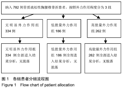
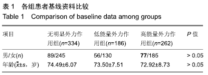
方法:回顾性分析2013年9月至2016年9月在广西中医药大学第一附属医院脊柱外科住院,明确诊断为骨质疏松性胸腰椎骨折的患者,3组中所有病例均行定量CT检查确诊为骨质疏松(骨密度值≤80 mg/cm3)。所有患者对治疗方案均知情同意,且得到医院伦理委员会批准。根据创伤史,分为无明显外力作用组(无明显诱因、无外力作用等)、低能量外力作用组(扭伤、弯腰提重物、扛重物等)、高能量外力作用组(平路摔倒臀部着地、跌倒、撞伤等),统计各组间性别、年龄、骨折部位(胸腰段和非胸腰段)、MRI STIR黑色线性信号椎体例数及所在的部位(胸腰段和非胸腰段)。3组间年龄属于计量资料采用方差分析;性别、骨折部位、MRI STIR黑色线性信号的椎体数量及所在部位属于计数资料,用Pearsonχ2检验。

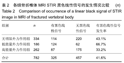
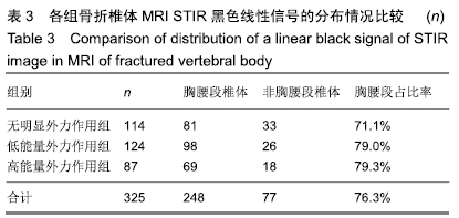
结果与结论:①纳入的3组病例共782例。无明显外力作用组334例,其中椎体内黑色线性信号114例;低能量外力作用组186例,其中椎体内黑色线性信号124例,高能量外力作用组262例,其中椎体内黑色线性信号87例;②3组间年龄、性别、骨折部位、MRI STIR黑色线性信号所在椎体部位差异均无显著性意义(P > 0.05);③3组间MRI STIR黑色线性信号比较差异有显著性意义(P < 0.05),分割P值,降低检验水准(α´=0.05/3=0.017)。低能量外力作用组分别与无明显外力作用组、高能量外力作用组比较,差异有显著性意义(P< 0.017);无明显外力作用组与高能量外力作用组比较,差异无显著性意义(P > 0.017);④并且低能量外力作用组中MRI STIR黑色线性信号出现率为66.7%,明显大于其他2组的43.1%和33.2%;⑤提示在创伤史中,相对于无明显外力作用和高能量外力作用,低能量外力作用的骨质疏松性胸腰椎骨折更容易导致MRI STIR黑色线性信号的出现,并且多见于胸腰段椎体。

ORCID: 0000-0003-4441-4355(钟远鸣)

中国组织工程研究杂志出版内容重点:人工关节;骨植入物;脊柱骨折;内固定;数字化骨科;组织工程


关键词: 骨质疏松, 胸腰椎骨折, MRI STIR黑色线性信号, 外力程度

Abstract:

BACKGROUND: With the aging of the society, the number of patients with osteoporotic vertebral fracture is increasing, mainly manifesting compression fracture of thoracolumbar body, which seriously affects the daily life of the elderly. Therefore, to study the relationship between the degree of external force and the performance of osteoporotic thoracolumbar body fracture on MRI STIR is to provide a better basis for clinical diagnosis and treatment.

OBJECTIVE: To explore the relationship between the size of external force and a linear black signal area of STIR image in MRI of thoracic and lumbar osteoporosis vertebral compression fractures.

METHODS: The hospitalized patients, who were diagnosed as thoracic and lumbar osteoporosis vertebral compression fractures, were retrospectively analyzed from September 2013 to September 2016 at the Department of Spine Surgery of The First Affiliated Hospital of Guangxi University of Chinese Medicine. All cases in the three groups were diagnosed as osteoporosis by quantitative CT (bone mineral density ≤ 80 mg/cm3). All patients signed the informed consent. This study was approved by the Hospital Ethics Committee. The patients were divided into three groups according to the different trauma history: Non-obvious external force group (without apparent cause or external force), low energy group (sprains, bent down to lift heavy objects, and carrying heavy items), high energy group (flat road down hips touchdown, falls, and bruise). Gender, age, fracture site (thoracic lumbar segment and non-thoracic lumbar segment), the number of the vertebrae and the position where would they occur with a linear black signal area of STIR image in MRI were analyzed in each group. Age was analyzed by analysis of variance. Gender, fracture site and the number of the vertebrae and the position were analyzed by Pearson chi-square test.

RESULTS AND CONCLUSION: (1) All the 782 cases were included in the three groups. There were 334 in the non-obvious external force group, which a linear black signal area of STIR image in MRI existed in 114 cases. There were 186 cases in low energy group, which a linear black signal area of STIR image in MRI existed in 124 cases. There were 262 cases in high energy group, which a linear black signal area of STIR image in MRI existed in 87 cases. (2) The age, gender, fracture site and the number of the vertebrae and the position in three groups were not statistically significantly different among the three groups (P > 0.05). (3) There were significant differences in a linear black signal area of STIR image in MRI among the three groups (P < 0.05) (α´=0.05/3=0.017). Significant differences were found as compared the low energy group with non-obvious external force group and high energy group (P < 0.017). No significant difference was determined between non-obvious external force group and high energy group (P > 0.017). (4) The occurrence rate of linear black signal area of STIR image in MRI was 66.7% and higher than other groups (43.1% and 33.2%). (5) In the history of trauma, low energy in external force has more opportunity to cause a linear black signal area of STIR image in MRI than non-obvious external force and high energy; and they often occur in thoracic and lumbar osteoporosis vertebrae.

Key words: osteoporosis, thoracolumbar compression fracture, linear black signals of MRI STIR, external force

中图分类号: