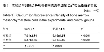
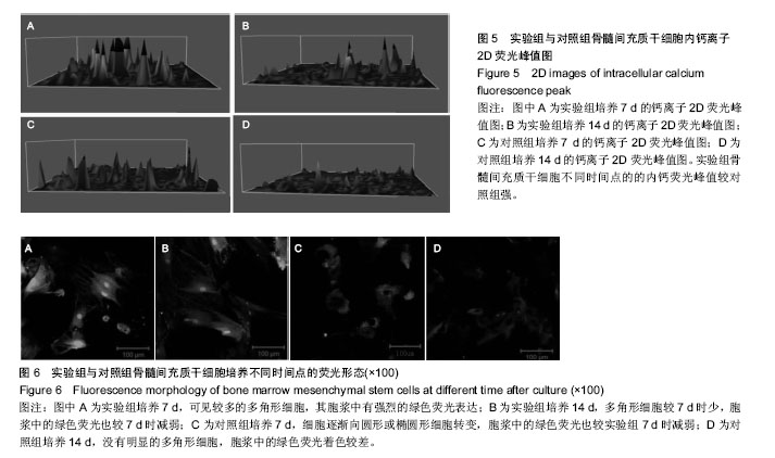
| [1] Pittenger MF,Mackay AM,Beck SC,et al.Multilineage potential of adult human mesenchymal stem cells.Science.1999; 284(5411):143-147. [2] Mao XZ,Zhou JN,Hu JZ,et al.Isolating and culturing Human bone marrow stromal cells in vitro and identifying tIle osteogenic activity.Chin J Modem Med. 2002;12(6):31-32.[3] Zhao ZP,Zhou JN,Li KH,et al.Multilineage potential of purified murine yolk sac mesenchymal stem cells in vitro.Chin J Modem Med.2002;12(18):25-27. [4] Rossi D,Sorrentino V.Molecular genetics of ryanodine receptors Ca2+-release channels.Cell Calcium. 2002; 32(5-6):307-319.[5] Borba VZ,Macas NC.The use of PTH in the treatment of osteoporosis.Arq Bras Endocrinol Metabol.2010;54(2):213-219.[6] Adams SL,Cohen AJ,Lassová L.Integration of signaling pathways regulating chondrocyte differentiation during endochondral bone formation.J Cell Physiol. 2007;213(3): 635-641.[7] The Ministry of Science and Technology of the People’s Republic of China. Guidance Suggestions for the Care and Use of Laboratory Animals.2006-09-30. [8] Uemura T,Dong J,Wang YC,et al.Transplantation of cultured bone cells using combinations of scaffolds and culture techniques.Biomaterials.2003;24:2277-2286.[9] Docheva D,Popov C,Mutschler W,et al.Human mesenchymal stem cells in contact with their environment: surface characteristics and the integrin system.J Cell Mol Med. 2007;11(1):21-38.[10] Ohgushi H,Capan AI.Stem cell technology and bioceramics: from cell to gene engineering.J Biomed Mater Res. 1999; 48(6):913-927. [11] Lei Z, Yongda L, Jun M, et al.Culture and neural differentiation of rat bone marrow mesenchymal stem cells in vitro.Cell Biol Int.2007;31(9):916-923.[12] Morikawa S,Mabuchi Y,Kubota Y,et al.Prospective identification, isolation, and systemic transplantation of multipotent mesenchymal stem cells in murine bone marrow.J Exp Med.2009;206(11):2483-2496.[13] Kode JA,Mukherjee S,Joglekar MV,et al.Mesenchymal stem cells: immunobiology and role in immunomodulation and tissue regeneration.Cytotherapy.2009;11(4):377-391.[14] Caplan AI.Mesenchymal stem cells.J Orthop Res. 1991;9: 641-650.[15] Di BG,Galderisi U,Fiorito C,et al.Dual role of parathyroid hormone in endothelial progenitor cells and marrow stromal mesenchymal stem cells.J Cell Physiol.2010; 222(2):474-480.[16] Mendez-Ferrer S,Michurina TV,Ferraro F,et al.Mesenchymal and haematopoietic stem cells form a unique bone marrow niche.Nature.2010;466(7308):829-834.[17] Geng S,Zhou S,Glowacki J.Age-related decline in osteoblastogenesis and 1alpha-hydroxylase/CYP27B1 in human mesenchymal stem cells: stimulation by parathyroid hormone.Aging Cell.2011;10(6):962-971.[18] Yu B,Zhao X,Yang C,et al.Parathyroid hormone induces differentiation of mesenchymal stromal/stem cells by enhancing bone morphogenetic protein signaling.J Bone Miner Res.2012;27(9):2001-2014.[19] Qiao GF,Zhou H,Li BY,et al.Antagonistic effects of berbamine on [Ca2+]i morbilization by KCL, norepinephrine, and caffeine in newborn rat cardiomycytes. Zhongguo Yao Li Xue Bao. 1999;20(4):292-296.[20] Hirose K,Kadowaki S,Tanabe M,et al.Spatiotemporal dynamics of inositol 1,4,5-trisphosphate that underlies complex Ca2+ mobilization patterns.Science. 1999;284(5419): 1527-1530.[21] Tfelt-Hansen J,MacLeod RJ,Chattopadhyay N,et al.Calciumsensing receptor stimulates PTHrP release by pathways dependent on PKC, p38 MAPK, JNK, and ERK1/2 in H-500 cells.Am J Physiol Endocrinol Metab.2003;285: E329-337.[22] Yano S,Macleod RJ,Chattopadhyay N,et al.Calcium-sensing receptor activation stimulates parathyroid hormone-related protein secretion in prostate cancer cells: role of epidermal growth factor receptor transactivation.Bone.2004;35:664-672. |
.jpg)

.jpg)