中国组织工程研究 ›› 2017, Vol. 21 ›› Issue (25): 4057-4061.doi: 10.3969/j.issn.2095-4344.2017.25.020

• 干细胞培养与分化 stem cell culture and differentiation • 上一篇    下一篇

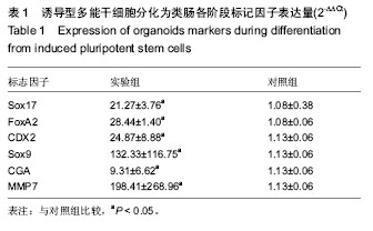
诱导型多能干细胞在体外三维环境中诱导分化出肠道类器官

李向阳1,赵 鑫1,相小松1,郑 鹏1,惠 璜2,嵇 武1   

  1. 1南京大学医学院附属金陵医院,解放军南京军区南京总医院解放军普通外科研究所,江苏省南京市  2100022解放军第二军医大学,上海市  200433
  • 修回日期:2017-04-14 出版日期:2017-09-08 发布日期:2017-10-09
  • 通讯作者: 嵇武,主任医师,教授,南京大学医学院附属金陵医院,解放军南京军区南京总医院解放军普通外科研究所,江苏省南京市 210002
  • 作者简介:李向阳,男,1989年生,安徽省亳州市人,汉族,南京大学在读硕士,主要从事肝胆胰腺外科研究。
  • 基金资助:

    南京军区医药卫生科研基金(15DX018)

Induced pluripotent stem cells differentiate into intestinal organoids in three-dimensional niche in vitro

Li Xiang-yang1, Zhao Xin1, Xiang Xiao-song1, Zheng Peng1, Hui Huang2, Ji Wu1   

  1. 1Jinling Hospital, Medical School of Nanjing University, PLA Research Institute of General Surgery, Nanjing General Hospital of Nanjing Military Region, Nanjing 210002, Jiangsu Province, China; 2Second Military Medical University of PLA, Shanghai 200433, China
  • Revised:2017-04-14 Online:2017-09-08 Published:2017-10-09
  • Contact: Ji Wu, Chief physician, Professor, Jinling Hospital, Medical School of Nanjing University, PLA Research Institute of General Surgery, Nanjing General Hospital of Nanjing Military Region, Nanjing 210002, Jiangsu Province, China
  • About author:Li Xiang-yang, Studying for master’s degree, Jinling Hospital, Medical School of Nanjing University, PLA Research Institute of General Surgery, Nanjing General Hospital of Nanjing Military Region, Nanjing 210002, Jiangsu Province, China
  • Supported by:

    the Medical Science Research Fund of Nanjing Military Region, No. 15DX018

摘要:

文章快速阅读:


文题释义:
肠道类器官:
肠道类器官是衍生于多能干细胞或肠道前体祖细胞,在细胞成分、特定功能和组织结构等方面与相应的器官高度相似,包含肠道隐窝结构及构成隐窝的干细胞、潘斯细胞、杯状细胞和内分泌细胞等。


摘要
背景:
诱导型多能干细胞是一类特殊的细胞,具有自我更新及多向分化潜力,在一定诱导条件下能够分化为肠道类器官。
目的:探究诱导型多能干细胞在体外特定条件下是否可分化出肠道类器官。
方法:复苏B6J小鼠18代诱导型多能干细胞,培养3 d克隆单位覆盖培养皿80%左右后,加入含有激活素A的培养基培养3 d,得到确定性内胚层,加入含成纤维细胞生长因子4和Wnt3A的培养基培养4 d,得到中后肠细胞球,将其与Matrigel培养基混合后滴入四孔板形成滴株样生长,在HEPES、Rspondin1、头蛋白、表皮细胞生长因子、B27添加剂等生长因子等共同作用下向肠道类器官分化,期间观察细胞形态变化,检测确定性内胚层FoxA2、Sox17表达,中后肠细胞球特异性标记物CDX2表达,肠道类器官特异性标记物 Sox9、CGA、MMP7表达。
结果与结论:①诱导型多能干细胞在含有激活素 A的培养基培养3 d,细胞初步分化,相互融合,确定性内胚层标记物FoxA2、Sox17表达较诱导型多能干细胞明显升高(P < 0.05);②确定性内胚层在含成纤维细胞生长因子4和WNT3A环境下培养第3天开始就有少量中后肠细胞球形成,第4天大量中后肠细胞球形成,中后肠细胞球标记物CDX2较确定性内胚层明显升高(P < 0.05);③中后肠细胞球培养3 d后逐渐形成肠道类器官,其特异性标记物 Sox9、CGA、MMP7表达明显高于中后肠细胞球(P < 0.05);④结果表明,诱导型多能干细胞可在体外三维环境中诱导分化出类肠道器官。

中国组织工程研究杂志出版内容重点:干细胞;骨髓干细胞;造血干细胞;脂肪干细胞;肿瘤干细胞;胚胎干细胞;脐带脐血干细胞;干细胞诱导;干细胞分化;组织工程
ORCID: 0000-0002-6058-2888(嵇武)

关键词: 干细胞, 分化, 诱导型多能干细胞, 定向分化, 体外培养, 确定性内胚层, 肠道类器官

Abstract:

BACKGROUND: Induced pluripotent stem cells (iPSCs) are a special type of cells with self-renewal and multi-differentiation potential, which can differentiate into intestinal organoids under certain conditions.
OBJECTIVE: To explore whether iPSCs can differentiate into intestinal organoids under specific conditions in vitro.
METHODS: iPSCs from B6J mice were recovered and cultured for 3 days until clone units covered about 80% of the culture dish, and then the cells were cultured in the medium containing Activin A for 3 days until the deterministic endoderm formed. Further, the culture medium was replaced by the medium with fibroblast growth factor 4 and Wnt3A for 4 days to differentiate into the spheroids with CDX2+. After that, spheroids were collected and mixed with Matrigel, and then the mixture was dropped into the 4-well plate and cultured with Rspondin1, Noggin, epidermal growth factor, B27 and other growth factors to differentiate into intestinal organoids. Cell morphology was observed, FoxA2 and Sox17 expresson in the deterministic endoderm was detected, and CDX2, Sox9, CGA, MMP7 were measured. 
RESULTS AND CONCLUSION: iPSCs were cultured with Activin A for 3 days with higher cell fusion, initial differentiation and FoxA2/Sox17 expression (P < 0.05) than those of non-induced iPSCs. Spheroids began to appear at the 3rd day after culture with fibroblast growth factor 4 and WNT3A, and formed a lot at the 4th day. And CDX2 expression in spheroids was significantly increased compared with that in the deterministic endoderm (P < 0.05). Organoids gradually formed after 3 days culture, which contained all cell types of intestinal organoids, and expressions of specific markers, Sox9, CGA, MMP7, were significantly higher than those in spheroids (P < 0.05). To conclude, iPSCs can be induced to differentiate into intestinal organoids in three-dimensional niche in vitro.

中国组织工程研究杂志出版内容重点:干细胞;骨髓干细胞;造血干细胞;脂肪干细胞;肿瘤干细胞;胚胎干细胞;脐带脐血干细胞;干细胞诱导;干细胞分化;组织工程

Key words: Stem Cells, Multipotent Stem Cells, Cell Differentiation, Tissue Engineering

中图分类号: