中国组织工程研究 ›› 2017, Vol. 21 ›› Issue (25): 3937-3942.doi: 10.3969/j.issn.2095-4344.2017.25.001

• 骨髓干细胞 bone marrow stem cells •    下一篇

成纤维细胞生长因子处理多次传代培养骨髓间充质干细胞的增殖与分化

宋明宇1,杨 勇2,吴 华2,王 蓉3   

  1. 华中科技大学同济医学院附属同济医院,1妇产科, 2骨科,湖北省武汉市 430030;3长江航运总医院消化内科,湖北省武汉市 430015
  • 修回日期:2017-03-26 出版日期:2017-09-08 发布日期:2017-10-09
  • 通讯作者: 王蓉,硕士,主治医师,长江航运总医院消化内科,湖北省武汉市 430015
  • 作者简介:宋明宇,男,1987年生,山东省潍坊市人,汉族,2015年华中科技大学毕业,博士,主治医师,主要从事骨髓间充质干细胞增殖分化方面研究。
  • 基金资助:

    国家自然科学基金青年基金项目(81301552,51707075)

Effects of fibroblast growth factor on proliferation and differentiation of serially passaged bone marrow mesenchymal stem cells

Song Ming-yu1, Yang Yong2, Wu Hua2, Wang Rong3   

  1. 1Department of Obstetrics and Gynecology, 2Department of Orthopedics, Tongji Hospital, Tongji Medical College, Huazhong University of Science and Technology, Wuhan 430030, Hubei Province, China; 3Department of Gastroenterology, General Hospital of the Yangtse River Shipping, Wuhan 430015, Hubei Province, China
  • Revised:2017-03-26 Online:2017-09-08 Published:2017-10-09
  • Contact: Wang Rong, Master, Attending physician, Department of Gastroenterology, General Hospital of the Yangtse River Shipping, Wuhan 430015, Hubei Province, China
  • About author:Song Ming-yu, M.D., Attending physician, Department of Obstetrics and Gynecology, Tongji Hospital, Tongji Medical College, Huazhong University of Science and Technology, Wuhan 430030, Hubei Province, China
  • Supported by:

    the National Natural Science Foundation of China for the Youth, No. 81301552, 51707075

摘要:

文章快速阅读:


文题释义:
成纤维细胞生长因子:
是一种分泌型的信号蛋白,有酸性(pI 5.6)和碱性(pI 9.6)2种,能促进成纤维细胞有丝分裂、中胚层细胞的生长,还可刺激血管形成,在细胞增殖、分化和伤口愈合中起着重要作用。
干细胞特性:干细胞具有两大特性:自我复制能力及多向分化能力。具体来说干细胞可以通过有丝分裂来产生与本身性质相同的子代干细胞,而且在一定的外界因素干预下,干细胞具有向多种特化细胞分化的能力。


摘要
背景:
骨髓间充质干细胞来源有限,而且在多次体外传代培养过程中,其细胞形态、增殖及多向分化能力会发生改变。
目的:探讨成纤维细胞生长因子对经过多次传代、细胞形态发生改变的骨髓间充质干细胞形态、增殖及多向分化能力的影响。
方法:①体外分离培养大鼠骨髓间充质干细胞,连续传代培养6次,观察其细胞形态变化;②将第6代细胞接种于96孔板中,随机分为对照组和成纤维细胞生长因子处理组,相应培养1,2,3,4,5,6,7 d后,用CCK-8试剂盒检测细胞增殖状况;③将第6代细胞接种于6孔板中,随机分为对照组和成纤维细胞生长因子处理组,分别用普通培养基和含成纤维细胞生长因子培养基培养7 d,换成骨、成脂、成软骨诱导培养基继续培养7 d,应用荧光定量PCR法检测成骨(RUNX2、ALP、OCN)、成脂(PPARγ2、AP2、ADIPOQ)、成软骨(SOX9、Collagen II、aggrecan)相关基因的表达,应用蛋白印记法检测RUNX2、PPARγ2、SOX9蛋白的表达;④将第6代细胞接种到6孔细胞培养板中,随机分为对照组及成纤维细胞生长因子处理组,分别用普通生长培养基及含成纤维细胞生长因子的生长培养基培养7 d,换用成骨、成脂、成软骨诱导培养基继续培养14 d,进行茜素红染色、油红O染色和阿利新蓝染色。
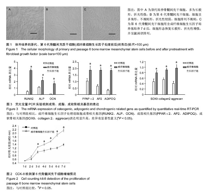
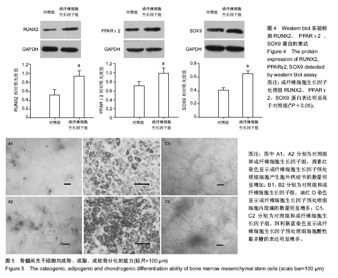
结果与结论:①经过连续6次传代培养,骨髓间充质干细胞形态发生明显变化,成纤维细胞生长因子处理7 d后其形态逐渐恢复原代特性;②与对照组相比,培养第3-7天成纤维细胞生长因子处理组的细胞增殖速度明显加快,差异有显著性意义(P < 0.05);③与对照组相比,成纤维细胞生长因子处理组细胞成骨相关基因(RUNX2,ALP,OCN)、成脂相关基因(PPARγ2,AP2,ADIPOQ)、成软骨相关基因(SOX9,collagen 2,aggrecan)表达明显升高,差异有显著性意义(P < 0.05);④成纤维细胞生长因子处理组RUNX2、PPARγ2、SOX9蛋白表达明显高于对照组(P < 0.05);⑤与对照组相比,成纤维细胞生长因子预处理组细胞产生胞外钙结节的数量、细胞内脂滴的数量、细胞酸性黏多糖的表达明显增加;⑥结果表明,成纤维细胞生长因子可以维持多次传代骨髓间充质干细胞的干细胞特性。


关键词: 干细胞, 骨髓干细胞, 骨髓间充质干细胞, 增殖, 分化, 细胞形态, 成骨, 成脂, 成软骨, 干细胞特性, 成纤维细胞生长因子, 国家自然科学基金

Abstract:

BACKGROUND: The source of bone marrow mesenchymal stem cells (BMSCs) is limited, and the cellular morphology, proliferation and multi-directional differentiation capacities can vary during serial passages in BMSCs in vitro.
OBJECTIVE: To study the effects of fibroblast growth factor (FGF) on cellular morphology, proliferation and differentiation of serially passaged BMSCs.
METHODS: (1) BMSCs were isolated from Sprague-Dawley rats and cultured. These cells were passaged six times in vitro, and the cellular morphology was observed and photographed. (2) BMSCs at passage 6 were seeded into 96-well plates and randomly divided into control group and FGF treatment group. The proliferation of cells in both groups was detected with cell counting kit-8 kit at days 1, 2, 3, 4, 5, 6, 7 after culture. (3) BMSCs at passage 6 were seeded into 6-well plates and randomly divided into control group and FGF treatment group. After 7 days treatment with growth medium or growth medium containing FGF, the cellular morphology was observed and photographed. And then the cells of both groups were treated with osteogenic induction medium, adipogenic induction medium and chondrogenic induction medium for the next 7 days. The osteogenic, adipogenic and chondrogenic related genes (RUNX2, ALP, OCN; PPARγ2, AP2, ADIPOQ; SOX9, collagen II, aggrecan) were detected with real-time PCR. The protein expressions of RUNX2, PPARγ2, SOX9 were detected with western blot assay. (4) BMSCs at passage 6 were seeded into 6-well plates and randomly divided into control group and FGF treatment group. After 7 days treatment with growth medium or growth medium containing FGF, the cells were cultured with osteogenic induction medium, adipogenic induction medium and chondrogenic induction medium for the next 14 days. Then, alizarin red S staining, oil red O staining and alcian blue staining were performed.
RESULTS AND CONCLUSION: (1) After in vitro passage for six times, the cellular morphology changed obviously, and FGF treatment recovered the characteristics of primary cells. (2) Compared with the control group, the cell proliferation in the FGF treatment group was significantly increased (P < 0.05). (3) Compared with the control group, the expression of osteogenic, adipogenic and chondrogenic related genes (RUNX2, ALP, OCN; PPARγ2, AP2, ADIPOQ; SOX9, collagen II, aggrecan) was increased significantly in the FGF treatment group (P < 0.05). The protein expressions of RUNX2, PPARγ2, SOX9 were also higher in the FGF treatment group than the control group (P < 0.05). (4) Compared with the control group, the number of extracellular calcium nodules, the number of intracellular lipid droplets, and the expression of acid acidic mucopolysaccharide were significantly increased after FGF pretreatment. To conclude, FGF pretreatment can preserve the stemness of BMSCs serially passaged in vitro.


Key words: Bone Marrow, Mesenchymal Stem Cells, Fibroblast Growth Factors, Cell Proliferation, Cell Differentiation, Tissue Engineering

中图分类号: