中国组织工程研究 ›› 2016, Vol. 20 ›› Issue (52): 7803-7808.doi: 10.3969/j.issn.2095-4344.2016.52.007

• 纳米生物材料 nanobiomaterials • 上一篇    下一篇

玻璃酸钠壳聚糖纳米粒对烧伤角膜新生血管生长的影响

鲁  静1,武士科2,陈  光1,赵  越1,李  丹1
  

  1. 河北大学附属医院,1眼科;2骨科,河北省保定市  071000
  • 收稿日期:2016-09-23 出版日期:2016-12-16 发布日期:2016-12-16
  • 通讯作者: 武士科,主治医师,河北大学附属医院骨科,河北省保定市 071000
  • 作者简介:鲁静,女,1981年生,汉族,河北省保定市人,硕士,主治医师,主要从事生物、细胞方面的研究。
  • 基金资助:

    河北省医学科学研究重点课题(zl20140018)

Effect of sodium hyaluronate/chitosan nanoparticles on the neovascularization in burned cornea

Lu Jing1, Wu Shi-ke2, Chen Guang1, Zhao Yue1, Li Dan1
  

  1. 1Department of Ophthalmology, 2Department of Orthopaedics, Affiliated Hospital of Hebei University, Baoding 071000, Hebei Province, China
  • Received:2016-09-23 Online:2016-12-16 Published:2016-12-16
  • Contact: Wu Shi-ke, Attending physician, Department of Orthopaedics, Affiliated Hospital of Hebei University, Baoding 071000, Hebei Province, China
  • About author:Lu Jing, Master, Attending physician, Department of Ophthalmology, Affiliated Hospital of Hebei University, Baoding 071000, Hebei Province, China
  • Supported by:

    the Key Subject of Medical Science Research of Hebei Province, No. zl20140018

摘要:

文章快速阅读:


文题释义:
玻璃酸钠:
又名透明质酸钠,是由N-乙酰葡萄糖醛酸反复交替而形成的一种高分子多糖体生物材料。玻璃酸钠为关节滑液的主要成分,是软骨基质的成分之一,在关节腔内起润滑作用,可覆盖和保护关节软骨,改善关节挛缩,抑制软骨变性变化表面,改善病理性关节液,增加滴滑功能。
玻璃酸钠壳聚糖纳米粒:将玻璃酸钠制作成由壳聚糖纳米粒包裹而成的药物,对体内外环境都有较强抵抗能力,纳米粒外层的聚电解质系统,能够延长蛋白质药物在外环境的保存期限,能够明显地提高治疗效果,并能降低药物带来的一定不良反应。

背景:将玻璃酸钠制作成由壳聚糖纳米粒包裹而成的药物,可以使药物更好地作用于烧伤角膜。
目的:验证玻璃酸钠壳聚糖纳米粒对烧伤角膜新生血管生长的影响。
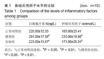
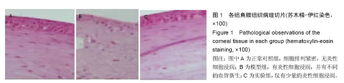
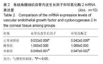
方法:将30只SD大鼠随机分为3组,模型组、实验组制备碱烧伤角膜模型,正常对照组不造模,实验组在造模后2周后滴注玻璃酸钠壳聚糖纳米粒悬液治疗,1次/d,10 μL/次,正常对照组与模型组给予等量生理盐水,持续4周。4周后,裂隙灯观察眼角膜新生血管动态生长情况;ELISA法测定白细胞介素6和肿瘤坏死因子α含量;苏木精-伊红染色观察角膜病理学变化;聚合酶链式反应实时荧光定量PCR检测检测血管内皮因子和环氧化酶2 mRNA表达量。
结果与结论:①与正常对照组比较,模型组角膜新生血管面积、白细胞介素6、肿瘤坏死因子α、血管内皮生长因子、环氧化酶2水平显著升高(P < 0.05,P < 0.01);与模型组比较,实验组角膜新生血管面积、白细胞介素6、肿瘤坏死因子α、血管内皮生长因子、环氧化酶2水平显著降低(P < 0.05);②模型组眼角组织有大量炎性细胞及新生血管产生,实验组仅有少量炎性细胞存在;③结果表明,玻璃酸钠壳聚糖纳米粒可抑制烧伤角膜新生血管的生长。

关键词: 生物材料, 纳米材料, 壳聚糖纳米材料, 玻璃酸钠, 烧伤角膜, 大鼠模型, 血管生长, 组织病理学, 白细胞介素6, 肿瘤坏死因子

Abstract:

BACKGROUND: Chitosan nanoparticles-encapsuled sodium hyaluronate is an effective drug for the burned cornea.
OBJECTIVE: To verify the effect of sodium hyaluronate/chitosan nanoparticles on the neovascularization in burned cornea.
METHODS: Thirty Sprague-Dawley rats were randomly divided into three groups, and the model of burned cornea caused by base was established in the rats of model and experimental groups, followed by respectively treated with 10 μL sodium hyaluronate/chitosan nanoparticle suspension and normal saline, once daily, for consecutive 4 weeks. Rats only given normal saline were used as controls. Four weeks later, the dynamic growth of newly formed blood vessels in the cornea was observed using silt lamp. The levels of tumor necrosis factor-α and interleukin-6 were detected by ELISA, histological changes of the cornea were observed by hematoxylin-eosin staining, and the mRNA expression levels of vascular endothelial growth factor and cyclooxygenase 2 were detected by real-time PCR.
RESULTS AND CONCLUSION: Compared with the control group, the area of the newly formed blood vessel and the levels of tumor necrosis factor-α, vascular endothelial growth factor and cyclooxygenase 2 were significantly increased in the model group (P < 0.05, P < 0.01). In the experimental group, all above indicators were significantly lower than those in the model group (P < 0.05). There were a large number of inflammatory cells and neovascularization in the model group, but only few inflammatory cells in the experimental group. These results show that sodium hyaluronate/chitosan nanoparticles can inhibit the neovascularization in the burned cornea.

Key words: Chitosan, Nanostructures, Cornea, Tissue Engineering

中图分类号: