中国组织工程研究 ›› 2016, Vol. 20 ›› Issue (42): 6284-6289.doi: 10.3969/j.issn.2095-4344.2016.42.008

• 软骨组织构建 cartilage tissue construction • 上一篇    下一篇

差速贴壁法原代培养椎间盘髓核细胞中的应用优势

胡永凯,孙浩林,漆龙涛,李淳德   

  1. 北京大学第一医院骨科,北京市  100034
  • 收稿日期:2016-08-02 出版日期:2016-10-14 发布日期:2016-10-14
  • 通讯作者: 李淳德,主任医师,北京大学第一医院骨科,北京市 100034
  • 作者简介:胡永凯,男,1988年生,湖南省邵阳市人,汉族,2016年北京大学毕业,硕士,医师,主要从事脊柱退行性疾病,脊柱畸形的研究。
  • 基金资助:

    国家自然科学基金(81441058)

Excellence of differential velocity adherent procedure in primary culture of nucleus pulposus cells of intervertebral disc

Hu Yong-kai, Sun Hao-lin, Qi Long-tao, Li Chun-de   

  1. Department of Orthopaedics, Peking University First Hospital, Beijing 100034, China
  • Received:2016-08-02 Online:2016-10-14 Published:2016-10-14
  • Contact: Li Chun-de, Chief physician, Department of Orthopaedics, Peking University First Hospital, Beijing 100034, Chinaa
  • About author:Hu Yong-kai, Master, Physician, Department of Orthopaedics, Peking University First Hospital, Beijing 100034, China
  • Supported by:

    the National Natural Science Foundation of China, No. 81441058

摘要:

文章快速阅读:

文题释义:
差速贴壁法:根据不同类型细胞黏附于某固相物质表面生长的能力的差异,即某些细胞黏附能力强或作用快而另一些细胞黏附能力弱或作用慢,接种后收集或去除未贴壁细胞,从而将某特定类型细胞进行分离以达到逐步纯化的目的。
原代培养:是指直接从机体取下组织、器官和细胞后立即进行培养。因此,较为严格地说是指成功传代之前的培养,此时的细胞保持原有细胞的基本性质。但通常把第1代至第10代以内的培养细胞统称为原代细胞培养。最常用的原代培养有组织块培养和分散细胞培养。
摘要
背景:
髓核细胞作为椎间盘的主要功能细胞是退变机制研究的重点对象,维持体外培养的髓核细胞的生理功能及细胞表型的稳定至关重要。
目的:通过对照研究差速贴壁法在大鼠椎间盘髓核细胞原代培养中的应用优势。
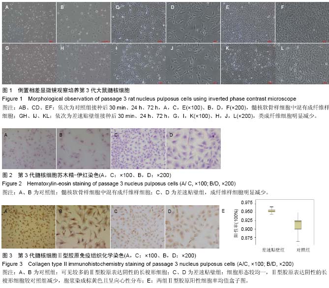
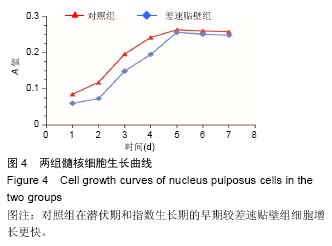
方法:取4周龄雄性Wistar大鼠20只,体外分离培养椎间盘髓核细胞,待原代细胞汇合近90%时进行分组传代,差速贴壁组在传代细胞贴壁30 min时,将未贴壁细胞吸出重新调整浓度后传至新的培养皿中,对照组不作处理。差速贴壁组第1、2代细胞均采用该法分离纯化。对两组第3代细胞进行细胞形态学比较及免疫组化鉴定纯度分析、CCK-8检测细胞功能活性并绘制细胞生长曲线。
结果与结论:①倒置显微镜观察及苏木精-伊红染色,差速贴壁组细胞均一性较对照组明显增高,对照组髓核细胞中混有较多的成纤维样细胞;②Ⅱ型胶原免疫组化鉴定及纯度分析,2组细胞胞浆均为被染成黄褐色,证实为髓核类软骨细胞;③纯度分析差速贴壁组阳性率显著高于对照组(P < 0.05);④CCK-8细胞生长曲线显示,2组细胞均经过2 d的生长潜伏期,3 d的指数生长期,进入生长停滞期,而对照组较差速贴壁组在潜伏期和指数增长期的早期生长更迅速。⑤结果说明,差速贴壁法是原代培养大鼠髓核细胞一种实用、有效的分离纯化方法;原代培养、经2次差速贴壁法分离纯化的第3代大鼠髓核细胞代谢旺盛、表型一致,细胞纯度更高,其对数增长期可以作为椎间盘细胞机制研究的最佳时期。

中国组织工程研究杂志出版内容重点:组织构建;骨细胞;软骨细胞;细胞培养;成纤维细胞;血管内皮细胞;骨质疏松组织工程
ORCID: 0000-0002-7368-1412(胡永凯)

关键词: 组织构建, 软骨组织工程, 差速贴壁, 髓核细胞, 纯化, 细胞去分化, 细胞老化, 国家自然科学基金

Abstract:

BACKGROUND: As the main function cell of intervertebral disc, nucleus pulposus cells are the focus of studying the degenerative mechanism; thereby, it is crucial to maintaining the physiological function of nucleus pulposus cells in vitro and the stability of the cell phenotype.
OBJECTIVE: To study the excellence of differential velocity adherent procedure in primary culture of nucleus pulposus cells of rat intervertebral disc through comparison.
METHODS: Twenty male Wistar rats aged 4 weeks were enrolled, and then nucleus pulposus cells of intervertebral disc were isolated and cultured in vitro; cell passage culture was performed in different groups when the primary cells were merged to 90%. Differential velocity adherent group cells adhered for 30 minutes, and non-adherent cells were aspirated and transferred to new culture dish after readjusting the concentration; the controls received no intervention. Passages 1 and 2 cells in the differential velocity adherent group were isolated and purified by the same procedure. The morphology of three generations of cells in the two groups was compared, the purity of the identification was detected by immunohistochemistry, the cell viability was detected by cell counting kit-8 and the cell growth curve was drawn.
RESULTS AND CONCLUSION: Inverted phase contrast microscope and hematoxylin-eosin staining revealed that the cell homogeneity of the differential velocity adherent group was significantly higher than that of the control group, and there were more kinds of fibroblast-like cells in nucleus pulposus cells in the control group. Identification and purity analysis of collagen type II showed that the cytoplasm of two groups were both stained brown, indicating that they were the nucleus pulposus chrondrocytes. The positive rate of differential velocity adherent group was significantly higher than that of the control group (P < 0.05). The cell growth curve of cell counting kit-8 showed cells in the two groups all passed by the latency phase within 2 days, then to the logarithmic phase of 3 days, and entered the lag phase, while the growth rate of the control group was more rapid during the latency and the early logarithmic phases.
These findings suggest that differential velocity adherence method is a practical and effective procedure for the isolation and purification of primary cultured rat nucleus pulposus cells. Through the primary culture, twice differential velocity adherence procedure, the passage 3 rat nucleus pulposus cells are metabolic exuberant, consistent with the phenotype, the cell purity is higher, and the logarithmic growth phase can be used as the optimal time for studying the mechanism of intervertebral disc cells.

中国组织工程研究杂志出版内容重点:组织构建;骨细胞;软骨细胞;细胞培养;成纤维细胞;血管内皮细胞;骨质疏松组织工程

Key words:  Intervetebral Disk, Cells, Cultured, Cell Aging, Tissue Engineering

中图分类号: