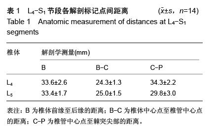
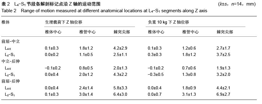
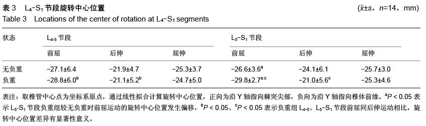
| [1] Alshami AM. Prevalence of spinal disorders and their relationships with age and gender. Saudi Med J. 2015; 36(6):725-730. [2] Tian W, Lv Y, Liu Y, et al. The high prevalence of symptomatic degenerative lumbar osteoarthritis in chinese adults: a population-based study. Spine (Phila Pa 1976). 2014;39(16):1301-1310. [3] Zhang SY, Huang P, Huang X, et al. Study on risk factors and predictive model for lumbar intervertebral disc herniation in the rural population. Zhonghua Liu Xing Bing Xue Za Zhi. 2009;30(11):1152-1155. [4] Mundt DJ, Kelsey JL, Golden AL, et al. An epidemiologic study of non-occupational lifting as a risk factor for herniated lumbar intervertebral disc. The Northeast Collaborative Group on Low Back Pain. Spine (Phila Pa 1976). 1993;18(5):595-602. [5] Taghipour-Darzi M, Ebrahimi-Takamjani E, Salavati M, et al. Construct validity of center of rotation in differentiating of lumbar segmental instability patients. J Back Musculoskelet Rehabil. 2014. [6] Gertzbein SD, Seligman J, Holtby R, et al. Centrode characteristics of the lumbar spine as a function of segmental instability. Clin Orthop Relat Res.1986; (208):48-51. [7] Schilling C, Pfeiffer M, Grupp TM, et al. The effect of design parameters of interspinous implants on kinematics and load bearing: an in vitro study. Eur Spine J. 2014;23(4):762-771. [8] Rousseau MA, Bradford DS, Bertagnoli R, et al. Disc arthroplasty design influences intervertebral kinematics and facet forces. Spine J. 2006;6(3): 258-266. [9] Yoshioka T, Tsuji H, Hirano N, et al. Motion characteristic of the normal lumbar spine in young adults: instantaneous axis of rotation and vertebral center motion analyses. J Spinal Disord. 1990;3(2): 103-113. [10] Schmidt H, Heuer F, Claes L, et al. The relation between the instantaneous center of rotation and facet joint forces-a finite element analysis. Clin Biomech (Bristol, Avon). 2008;23(3):270-278. [11] Schmidt H, Midderhoff S, Adkins K, et al. The effect of different design concepts in lumbar total disc arthroplasty on the range of motion, facet joint forces and instantaneous center of rotation of a L4-5 segment. Eur Spine J. 2009;18(11):1695-1705. [12] Gertzbein SD, Holtby R, Tile M, et al. Determination of a locus of instantaneous centers of rotation of the lumbar disc by moire fringes: a new technique. Spine (Phila Pa 1976). 1984;9(4):409-413. [13] Zander T, Rohlmann A, Bergmann G. Influence of different artificial disc kinematics on spine biomechanics. Clin Biomech (Bristol, Avon). 2009; 24(2):135-142. [14] Bogduk N, Amevo B, Pearcy M. A Biological basis for instantaneous centres of rotation of the vertebral column. Proc Inst Mech Eng H. 1995;209(3):177-183. [15] Xia Q, Wang S, Kozanek M, et al. In-vivo motion characteristics of lumbar vertebrae in sagittal and transverse planes. J Biomech. 2010;43(10): 1905-1909. [16] 胥鸿达,苗军,夏群.生理载荷下腰椎退变滑脱椎管容积的体内三维试验[J].中国组织工程研究,2014,18(53): 8634-8640.[17] Miao J, Wang S, Wan Z, et al. Motion Characteristics of the Vertebral Segments with Lumbar Degenerative Spondylolisthesis in Elderly Patients.Eur Spine J. 2013; 22(2):425-431.[18] 王博韬,夏群,苗军,等.应用数字骨科技术观测腰椎失稳节段间在体三维运动特点[J].中华医学杂志,2014,94(29): 2264-2268.[19] Wang S, Passias P, Li G, et al. Measurement of vertebral kinematics using noninvasive image matching method-validation and application. Spine (Phila Pa 1976). 2008;33(11):E355-E361. [20] Wang S, Park WM, Gadikota HR, et al. A combined numerical and experimental technique for estimation of the forces and moments in the lumbar intervertebral disc. Comput Methods Biomech Biomed Engin. 2013; 16(12):1278-1286. [21] Yao Q, Wang S, Shin JH, et al. Motion characteristics of the lumbar spinous processes with degenerative disc disease and degenerative spondylolisthesis. Eur Spine J. 2013;22(12):2702-2709. [22] Yao Q, Wang S, Shin JH, et al. Lumbar Facet Joint Motion in Patients with Degenerative Spondylolisthesis. J Spinal Disord Tech. 2013;26(1):E19-E27. [23] Pearcy MJ, Bogduk N. Instantaneous Axes of Rotation of the Lumbar Intervertebral Joints. Spine (Phila Pa 1976). 1988;13(9):1033-1041. [24] Samagh Sanjum P, Rosen Charles D, Otarodifard Karimdad, et al. New method for determining apparent axial center of rotation of lumbar and thoracic spine segments. J Rehabil Res Dev. 2011;48(5):587. [25] Li G, Wang S, Passias P, et al. Segmental in vivo vertebral motion during functional human lumbar spine activities. Eur Spine J. 2009;18(7):1013-1021. [26] Wang S, Park WM, Kim YH, et al. In Vivo Loads in the Lumbar L3-4 Disc During a Weight Lifting Extension. Clin Biomech (Bristol, Avon). 2014;29(2):155-160. [27] Goel VK, Monroe BT, Gilbertson LG, et al. Interlaminar shear stresses and laminae separation in a disc. finite element analysis of the L3-L4 motion segment subjected to axial compressive loads. Spine (Phila Pa 1976). 1995;20(6):689-698. [28] An KN, Chao EY. Kinematic analysis of human movement. Ann Biomed Eng. 1984;12(6): 585-597. [29] Anderst W, Baillargeon E, Donaldson W, et al. Motion path of the instant center of rotation in the cervical spine during in vivo dynamic flexion-extension: implications for artificial disc design and evaluation of motion quality after arthrodesis. Spine (Phila Pa 1976). 2013;38(10):E594-E601. [30] Alapan Y, Sezer S, Demir C, et al. Load sharing in lumbar spinal segment as a function of location of center of rotation. J Neurosurg Spine. 2014;20(5): 542-549. |
.jpg)
.jpg)
.jpg)
.jpg)