中国组织工程研究 ›› 2015, Vol. 19 ›› Issue (52): 8401-8405.doi: 10.3969/j.issn.2095-4344.2015.52.007

• 组织工程骨及软骨材料 tissue-engineered bone and cartilage materials • 上一篇    下一篇

硼硅酸盐对成骨细胞体外生物活性的影响

程中华,薛 威,王李琴,黄清芳,吴成欢,桂凯红,黄 林,蔡 莹,韩艳芳,蒋彩霞   

  1. 黄冈市中心医院骨科,湖北省黄冈市 438000
  • 收稿日期:2015-09-21 出版日期:2015-12-17 发布日期:2015-12-17
  • 通讯作者: 黄林,硕士,黄冈市中心医院骨科,湖北省黄冈市 438000
  • 作者简介:程中华,男,1969年生,湖北省武汉市人,汉族,2003年湖北中医学院毕业,硕士,主任医师, 现任黄冈市骨科学会主委,长江大学医学院附属黄冈市中心医院骨科主任,主要从事骨与关节研究。
  • 基金资助:

    黄冈市科学技术研究与开发计划项目(s2015-01-0130084)

Borosilicate effect on in vitro biological activity of osteoblasts 

Cheng Zhong-hua, Xue Wei, Wang Li-qin, Huang Qing-fang, Wu Cheng-huan, Gui Kai-hong, Huang Lin, Cai Ying, Han Yan-fang, Jiang Cai-xia   

  1. Department of Orthopedics, Huanggang Central Hospital, Huanggang 438000, Hubei Province, China
  • Received:2015-09-21 Online:2015-12-17 Published:2015-12-17
  • Contact: Huang Lin, Master, Department of Orthopedics, Huanggang Central Hospital, Huanggang 438000, Hubei Province, China
  • About author:Cheng Zhong-hua, Master, Chief physician, Department of Orthopedics, Huanggang Central Hospital, Huanggang 438000, Hubei Province, China
  • Supported by:

    the Science and Technology Research and Development Program of Huanggang City of China, No. s2015-01-0130084

摘要:

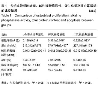
背景:硼硅酸盐不仅可通过矿化作用形成羟基碳酸盐磷灰石层,而且具有强化学反应活性,可促进骨细胞再生。
目的:通过体外培养实验观察硼硅酸盐生物玻璃对兔成骨细胞生长行为的影响。
方法:根据 ISO10993-12:2007 的要求制备硼硅酸盐生物玻璃初次浸提液与二次浸提液。分离培养兔骨髓间充质干细胞,取第2代细胞诱导生成成骨细胞。取第5-15代成骨细胞,分别以硼硅酸盐生物玻璃初次浸提液、硼硅酸盐生物玻璃二次浸提液与α-MEM培养基培养,观察硼硅酸盐生物活性玻璃对成骨细胞增殖、蛋白合成、碱性磷酸酶活性、细胞凋亡及细胞横向与纵向迁移的影响。
结果与结论:初次浸提液组与二次浸提液组成骨细胞增殖优于α-MEM培养基组(P < 0.05),且初次浸提液组成骨细胞增殖优于二次浸提液组(P < 0.05)。初次浸提液组成骨细胞总蛋白含量高于二次浸提液组与α-MEM培养基组(P < 0.05)。3组间成骨细胞碱性磷酸酶活性、凋亡率、横向迁移距离及Transwell 中穿膜细胞数比较差异均无显著性意义。表明硼硅酸盐生物玻璃具有良好的细胞相容性,对成骨细胞增殖有一定的良性调节作用。
 

关键词: 生物材料, 骨生物材料, 硼硅酸盐, 骨髓间充质干细胞, 成骨细胞, 碱性磷酸酶, 增殖

Abstract:

BACKGROUND: Borosilicate cannot only be mineralized to form hydroxy carbonate apatite layer, but also have strong chemical reactivity to promote bone cell regeneration.
OBJECTIVE: To investigate the effect of the borosilicate bioglass on the growth behavior of rabbit osteoblasts through in vitro culture experiment.
METHODS: The initial and secondary extracts of borosilicate bioglass were prepared according to the requirement of ISO10993-12: 2007. The bone marrow mesenchymal stem cells of rabbits were isolated and cultured. The second generation bone marrow mesenchymal stem cells were induced to differentiate into osteoblasts. The osteoblasts of the 5th-15th generations were obtained and cultured with the initial and secondary extracts of borosilicate bioglass and α-MEM medium, respectively. The effects of borosilicate bioglass on the osteoblasts proliferation, protein synthesis, alkaline phosphatase activity, cell apoptosis, and cell migration in horizontal and vertical direction were observed.
RESULTS AND CONCLUSION: The osteoblasts proliferation in the initial extract and secondary extract groups was better than that in the α-MEM medium group (P < 0.05). The osteoblasts proliferation in the initial extract group was better than that in the secondary extract group (P < 0.05). The total protein content of osteoblasts in the initial extract group was higher than that in the secondary extract and α-MEM medium group (P < 0.05). There were no significant differences in the alkaline phosphatase activity, apoptosis rate, horizontal migration distance of osteoblast and transmembrane cell number in Transwell between these three groups. These results demonstrate that borosilicate bioglass has good biocompatibility and has a certain benign regulatory role in  osteoblast proliferation.
 

中图分类号: