| [1] 张文文.用于荧光免疫分析的稀土掺杂无机发光纳米粒子的研究[D].广东:暨南大学,2007.
[2] Jiang G,Pichaandi J,JohnsonNJ,et al.An effective polymer cross-Linking strategy to obtain stable dispersions of upconverting NaYF4 nanoparticles in buffers and biological growth media for biolabeling applications. Langmuir. 2012; 28(6):3239-3247.
[3] Wang ZL,Hao J,Chan HL,et al.Simultaneous synthesis and functionalization of water-soluble up-conversion nanoparticles for in-vitro cell and nude mouse imaging.Nanoscale. 2011; 3(5): 2175-2181.
[4] Zhan Q,Qian J,Liang H,et al.Using 915nm laser excited Tm3+/ Er3+/ Ho3+-doped NaYbF4 unconversion nanoparticles for in vitro and deeper in vivo bioimaging without overheating irradiation.ACS Nano.2011;5(5): 3744-3757.
[5] Williams DF.On the mechanisms of biocompatibility. Biomaterials.2008;29(20): 2941-2953.
[6] 国家医疗器械生物学评价标准化技术委员会.GB-T16886 15-2003:医疗器械生物学评价[M].北京:中国标准出版社, 2003: 120-127.
[7] 李瑞,王青山.生物材料生物相容性的评价方法和发展趋势[J].中国组织工程研究, 2011,15(29):5471-5474.
[8] Leevy WM,Gammon ST,Jiang H,et al.Optical imaging of bacterial infection in living mice using a fluorescent near-infrared molecular probe.Journal of the American Chemical Society.2006;128(51):16476-16477.
[9] Zhang M, Yu M, Li F, et al. A highly selective fluorescence turn-on sensor for cysteine/homocysteine and its application in bioimaging.J Am Chem Soc. 2006;128(51):16476-16477.
[10] Xiong L,Yu M,Cheng M,et al.A photostable fluorescent probe for targeted imaging of tumour cells possessing integrin αvβ3. Mol Biosyst.2009;5(3): 241-243.
[11] [Bruchez M,Moronne M,Gin P,et al.Semiconductor nanocrystals as fluorescent biological labels. Science. 1998; 281(5385):2013-2016.
[12] Xiong LQ,Chen ZG,Yu MX,et al.Synthesis, characterization, and in vivo targeted imaging of amine-functionalized rare-earth up-converting nanophosphors. Biomaterials. 2009; 30(29):5592-5600.
[13] Smith AM,Duan H,Mohs AM,et al.Bioconjugated quantum dots for <i> in vivo </i> molecular and cellular imaging.Adv Drug Deliv Rev.2008;60(11):1226-1240.
[14] Wang X,Ren X,Kahen K,et al.Non-blinking semiconductor nanocrystals.Nature. 2009;459(7247):686-689.
[15] Efros AL.Nanocrystals: Almost always bright.Nat Mater. 2008;7(8):612-613.
[16] Mahler B,Spinicelli P,Buil S,et al.Towards non-blinking colloidal quantum dots.Nat Mater.2008;7(8):659-664.
[17] Derfus AM,Chan WCW,Bhatia SN.Probing the cytotoxicity of semiconductor quantum dots.Nano Lett.2004;4(1):11-18.
[18] Hardman R.A toxicologic review of quantum dots: toxicity depends on physicochemical and environmental factors.Environ Health Perspect. 2006;114(2):165-172.
[19] Shen J,Sun LD,Yan CH.Luminescent rare earth nanomaterials for bioprobe applications.Dalton Trans. 2008;(42):5687-5697.
[20] Vetrone F,Capobianco JA. Lanthanide-doped fluoride nanoparticles: luminescence, upconversion, and biological applications.IntJNanotechnol.2008;5(9):1306-1339.
[21] Dosev D,Nichkova M,Kennedy IM.Inorganic lanthanide nanophosphors in biotechnology.J Nanosci Nanotechnol. 2008;8(3):1052-1067.
[22] Escribano P,Julián-López B,Planelles-Aragó J,et al.Photonic and nanobiophotonic properties of luminescent lanthanide- doped hybrid organic–inorganic materials. J Mater Chem. 2008;18(1):23-40.
[23] Bouzigues C, Gacoin T, Alexandrou A. Biological applications of rare-earth based nanoparticles.Acs Nano. 2011;5(11): 8488-8505.
[24] Näreoja T,Vehniäinen M,Lamminmäki U,et al.Study on nonspecificity of an immuoassay using Eu-doped polystyrene nanoparticle labels.J Immunol Methods. 2009;345(1):80-89.
[25] Bruchez M,Moronne M,Gin P,et al.Semiconductor nanocrystals as fluorescent biological labels.Science. 1998; 281(5385):2013-2016.
[26] Qian J,Wang Y,Gao X,et al.Carboxyl-functionalized and bio-conjugated silica-coated quantum dots as targeting probes for cell imaging. J Nanosci Nanotechnol. 2010;10(3): 1668-1675.
[27] Smith JE,Wang L,Tan W.Bioconjugated silica-coated nanoparticles for bioseparation and bioanalysis.Trends Anal Chem.2006;25(9):848-855.
[28] Wang C,Ma Z,Wang T,et al.Synthesis, Assembly, and Biofunctionalization of Silica-Coated Gold Nanorods for Colorimetric Biosensing.Adv Funct Mater. 2006;16(13): 1673-1678.
[29] Hoshino A,Fujioka K,Oku T,et al.Physicochemical properties and cellular toxicity of nanocrystal quantum dots depend on their surface modification. Nano Lett.2004;4(11): 2163-2169.
[30] Li Z,Zhang Y.Monodisperse Silica-Coated Polyvinylpyrrolidone/NaYF4 Nanocrystals with Multicolor Upconversion Fluorescence Emission.Angewandte Chemie. 2006;118(46):7896-7899.
[31] Xiong LQ,Chen ZG,Yu MX,et al.Synthesis, characterization, and <i> in vivo </i> targeted imaging of amine-functionalized rare-earth up-converting nanophosphors.Biomaterials. 2009; 30(29):5592-5600.
[32] Zhou JC,Yang ZL,Dong W,et al.Bioimaging and toxicity assessments of near-infrared upconversion luminescent NaYF< sub> 4</sub>: Yb, Tm nanocrystals.Biomaterials. 2011;32(34):9059-9067. |
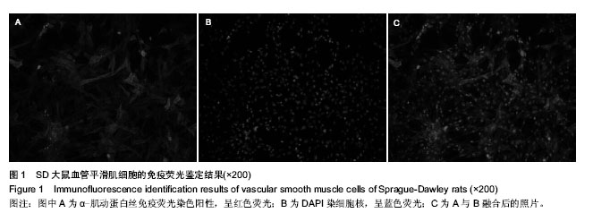
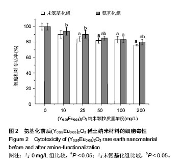
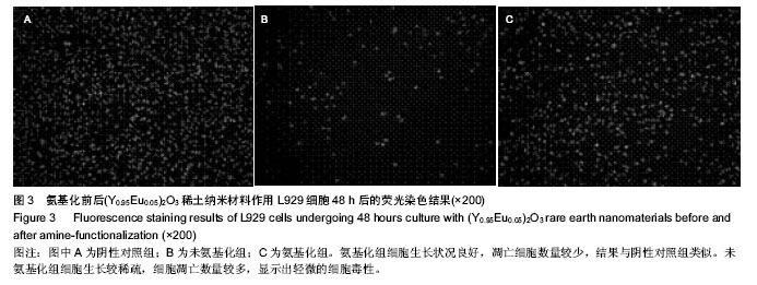
.jpg)