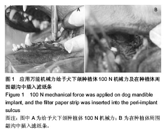
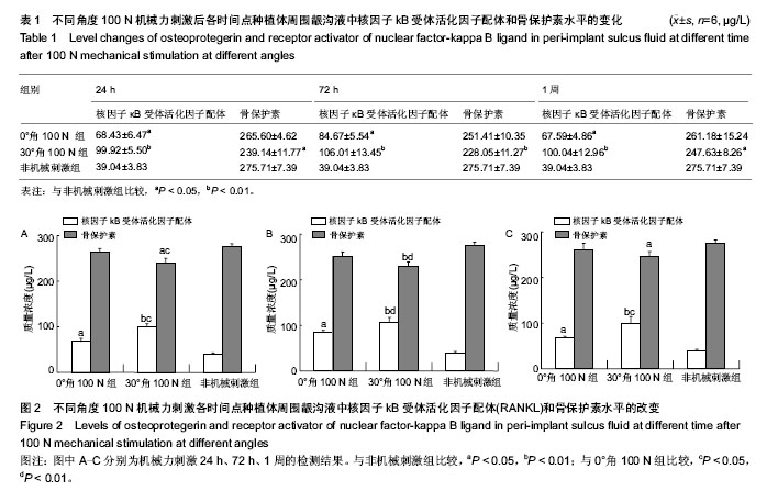
| [1] Di P, Lin Y, Li JH,et al.The All-on-Four implant therapy protocol in the management of edentulous Chinese patients. Int J Prosthodont. 2013;26(6):509-516.
[2] Maló P,Nobre MD,Lopes A. Immediate loading of 'All-on-4' maxillary prostheses using trans-sinus tilted implants without sinus bone grafting: a retrospective study reporting the 3-year outcome.Eur J Oral Implantol.2013;6(3): 273-283.
[3] Jensen OT.Complete arch site classification for all-on-4 immediate function. J Prosthet Dent.2014;112(4): 741-751.
[4] Ayna M,Gülses A,Açil Y.Comprehensive comparison of the 5-year results of 'All-on-4™' mandibular implant systems with acrylic and ceramic suprastructures, respectively. J Oral Implantol. 2014.[Epub ahead of print]
[5] Taruna M,Chittaranjan B,Sudheer N,et al.Prosthodontic perspective to all-on-4® concept for dental implants.J Clin Diagn Res.2014;8(10):ZE16-19.
[6] 中华人民共和国科学技术部.关于善待实验动物的指导性意见. 2006-09-30.
[7] Brunski JB.Biomechanical aspects of the optimal number of implants to carry a cross-arch full restoration.Eur J Oral Implantol. 2014;7 Suppl 2:S111-131.
[8] Tian K,Chen J,Han L,et al.Angled abutments result in increased or decreased stress on surrounding bone of single-unit dental implants: a finite element analysis. Med Eng Phys.2012;34(10): 1526-1531.
[9] Paul S,Padmanabhan TV,Swarup S,et al.Comparison of strain generated in bone by "platform-switched" and "non-platform-switched" implants with straight and angulated abutments under vertical and angulated load: a finite element analysis study.Indian J Dent Res.2013;24(1): 8-13.
[10] Kumar GA,Mahesh B,George D.Three Dimensional Finite Element Analysis of Stress Distribution Around Implant with Straight and Angled Abutments in Different Bone Qualities.J Indian Prosthodont Soc.2013;13(4):466-472.
[11] Ogawa T,Vandamme K,Zhang X,et al.Stimulation of titanium implant osseointegration through high-frequency vibration loading is enhanced when applied at high acceleration.Calcif Tissue Int.2014;95(5):467-475.
[12] Zhang X,Duyck J,Vandamme K,et al.Ultrastructural characterization of the implant interface response to loading.J Dent Res.2014;93(3):313-318.
[13] Browaeys H,Dierens M,Ruyffelaert C,et al.Ongoing Crestal Bone Loss around Implants Subjected to Computer-Guided Flapless Surgery and Immediate Loading Using the All-on-4 Concept. Clin Implant Dent Relat Res.2014.doi: 10.1111/cid.1219[Epub ahead of print]
[14] Arun Kumar G,Mahesh B,George D.Three Dimensional Finite Element Analysis of Stress Distribution Around Implant with Straight and Angled Abutments in Different Bone Qualities. J Indian Prosthodont Soc.2013;13(4):466-472.
[15] Canullo L,Coelho PG, Bonfante EA,et al.Angled abutments result in increased or decreased stress on surrounding bone of single-unit dental implants: a finite element analysis. Med Eng Phys.2012;34(10):1526-1531.
[16] Araújo AA ,Varela H,Brito GA,et al.Azilsartan increases levels of IL-10, down-regulates MMP-2, MMP-9, RANKL/RANK, Cathepsin K and up-regulates OPG in an experimental periodontitis model.PLoS One.2014;9(5):e96750.
[17] Nogueira AV,Nokhbehsaim M,Eick S,et al.Biomechanical Loading Modulates Proinflammatory and Bone Resorptive Mediators in Bacterial-Stimulated PDL Cells. Mediators Inflamm.2014;2014:425421.
[18] Repeke CE,Cardoso CR,Claudino M,et al.Non-inflammatory destructive periodontal disease: a clinical, microbiological, immunological and genetic investigation. J Appl Oral Sci. 2012;20(1):113-121.
[19] Belibasakis GN,Bostanci N.The RANKL-OPG system in clinical periodontology. J Clin Periodontol.2012;39:239-248.
[20] Sexton WM,Lin Y,Kryscio RJ,et al.Salivary biomarkers of periodontal disease in response to treatment.J Clin Periodontol. 2011;38:434-441.
[21] Buduneli N,Kinane DF.Host-derived diagnostic markers related to soft tissue destruction and bone degradation in periodontitis.J Clin Periodontol. 2011;38(Suppl 11):85-105.
[22] Costa PP,Trevisan GL,Macedo GO,et al.Salivary interleukin-6, matrix metalloproteinase-8, and osteoprotegerin in patients with periodontitis and diabetes. J Periodontol. 2010;81: 384-391.
[23] Al-Sabbagh M,Alladah A,Lin Y,et al.Bone remodeling- associated salivary biomarker MIP-1α distinguishes periodontal disease from health. J Periodontal Res. 2012; 47(3): 389-395.
[24] Tobón-Arroyave SI,Isaza-Guzmán DM,Restrepo-Cadavid EM,et al.Association of salivary levels of the bone remodelling regulators sRANKL and OPG with periodontal clinical status.J Clin Periodontol.2012;39(12):1132-1140.
[25] Tabari ZA, Azadmehr A,Tabrizi MAA,et al.Salivary soluble receptor activator of nuclear factor kappa B ligand/osteoprotegerin ratio in periodontal disease and health. J Periodontal Implant Sci.2013;43(5):227-232.
[26] Lin D,Li L,Sun Y,et al.IL-17 regulates the expressions of RANKL and OPG in human periodontal ligament cells via TRAF6/TBK1-JNK/NF-κB pathways.Immunology. 2014.doi: 10.1111/imm.12395. [Epub ahead of print]
[27] Prates TP,Taira TM,Holanda MC,et al.NOD2 contributes to Porphyromonas gingivalis-induced bone resorption.J Dent Res.2014;93(11):1155-1162.
[28] Araújo AA,Souza TO,Moura LM,et al.Effect of telmisartan on levels of IL-1, TNF-α, down-regulated COX-2, MMP-2, MMP-9 and RANKL/RANK in an experimental periodontitis model.J Clin Periodontol.2013;40(12):1104-1111.
[29] Takahashi S,Fukuda M,Mitani A,et al. Follicular dendritic cell-secreted protein is decreased in experimental periodontitis concurrently with the increase of interleukin-17 expression and the Rankl/Opg mRNA ratio.J Periodontal Res. 2014;49(3):390-397.
[30] Sarlati F,Sattari M,Razzaghi S,et al.Receptor activator of nuclear factor kappa B ligand and osteoprotegerin levels in gingival crevicular fluid. Dent Res J (Isfahan). 2012;9(6): 752-757.
[31] Bostanci N,Saygan B,Emingil G,et al.Effect of periodontal treatment on receptor activator of NF-κB ligand and osteoprotegerin levels and relative ratio in gingival crevicular fluid.J Clin Periodontol.2011;38:428-433.
[32] Petcu CM,Nitoi D,Mercut V,et al. Masticatory tensile developed in upper anterior teeth with chronic apical periodontitis. A finite-element analysis study. Rom J Morphol Embryol.2013;54(3):587-592.
[33] 王稚英,李世德,马晓凜,等.个性化钛板与纳米羟基磷灰石及自体骨修复兔上颌骨缺损.中国组织工程研究与临床康复,2010, 14(16):2851-2854. |