中国组织工程研究 ›› 2015, Vol. 19 ›› Issue (5): 681-684.doi: 10.3969/j.issn.2095-4344.2015.05.005

• 器官移植动物模型 organ transplantation and animal model • 上一篇    下一篇

原代缺氧缺糖损伤神经元模型Cdh1及其下游底物的表达

钱  巍,邱  瑾,祁月红,姚文龙,张  雪,张传汉   

  1. 华中科技大学同济医学院附属同济医院麻醉学教研室,湖北省武汉市  430030
  • 修回日期:2014-11-17 出版日期:2015-01-30 发布日期:2015-03-02
  • 通讯作者: 张传汉,华中科技大学同济医学院附属同济医院麻醉学教研室,湖北省武汉市 430030
  • 作者简介:钱巍,华中科技大学同济医学院附属同济医院麻醉学教研室,湖北省武汉市 430030
  • 基金资助:

    国家自然科学基金资助项目(30571788)

Expression of Cdh1 and its downstream substrates in primary neurons after oxygen-glucose deprivation

Qian Wei, Qiu Jin, Qi Yue-hong, Yao Wen-long, Zhang Xue, Zhang Chuan-han   

  1. Department of Anesthesiology, Tongji Hospital of Tongji Medical College, Huazhong University of Science and Technology, Wuhan 430030, Hubei Province, China
  • Revised:2014-11-17 Online:2015-01-30 Published:2015-03-02
  • Contact: Zhang Chuan-han, Department of Anesthesiology, Tongji Hospital of Tongji Medical College, Huazhong University of Science and Technology, Wuhan 430030, Hubei Province, China
  • About author:Qian Wei, Department of Anesthesiology, Tongji Hospital of Tongji Medical College, Huazhong University of Science and Technology, Wuhan 430030, Hubei Province, China
  • Supported by:

    the National Natural Science Foundation of China, No. 30571788

摘要:

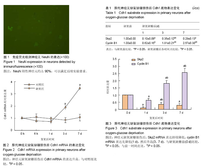
背景:课题组前期实验已证实Cdh1在大鼠海马、皮质均有大量表达,且体外实验发现神经元中Cdh1表达高于神经干细胞,可能与神经干细胞向神经元分化有关。但细胞周期末期促进复合物调节亚基Cdh1在缺血性神经元损伤中的作用,尚不明确。
目的:体外构建原代缺氧缺糖损伤神经元模型,观察Cdh1及其下游底物表达变化。
方法:取出生24 h内乳鼠大脑皮质,体外培养原代神经元并通过免疫荧光染色进行鉴定。使用无糖Earle’s 液替代细胞培养液,利用三气培养箱充以氮气建立原代神经元缺氧缺糖模型,缺氧处理1 h后复氧。于缺氧前、缺氧缺糖损伤后6 h、1 d,3 d,7 d采用实时荧光定量PCR检测神经元Cdh1及其下游底物Skp2、Cyclin B1的表达。
结果与结论:体外缺氧缺糖损伤后,原代神经元Cdh1及其下游底物Cyclin B1表达上调(P < 0.05),Skp2表达均下调(P < 0.05)。提示,体外缺氧缺糖损伤后神经元Cdh1表达升高,可能通过泛素化降解Skp2参与缺氧性神经元凋亡等病理过程。


中国组织工程研究
杂志出版内容重点:肾移植肝移植移植;心脏移植;组织移植;皮肤移植;皮瓣移植;血管移植;器官移植组织工程


全文链接:

关键词: 实验动物, 脑及脊髓损伤模型, 细胞周期末期促进复合物, Cdh1, 神经元, 缺氧, 缺糖, 损伤, 下游底物, Skp2, 国家自然科学基金

Abstract:

BACKGROUND: Cdh1 has been shown to express in rat hippocampus and cortex in a large number. Moreover, in vitro test demonstrated that Cdh1 expression was higher in neurons than in neural stem cells, which possibly associated with the differentiation of neural stem cells into neurons. However, the effects of anaphase promoting complex Cdh1 on ischemic neuronal damage remain unclear.
OBJECTIVE: To investigate the expression of Cdh1 and its downstream substrate in primary cultured neurons with oxygen-glucose deprivation.
METHODS: Primary neurons from cortex of postnatal 24-hour rat pups were cultured in vitro, and identified by immunofluorescence staining. The oxygen- and glucose-deprived models were established by three gas incubator filled with nitrogen in sugar-free Earle’s solution. After 1 hour of hypoxia, reoxygenation was conducted. Real-time fluorescent quantitative PCR was used to detect the mRNA expression of Cdh1 and its downstream substrates Skp2, Cyclin B1 before hypoxia, 6 hours, 1, 3, 7 days after oxygen glucose deprivation.
RESULTS AND CONCLUSION: After oxygen glucose deprivation, the expression of Cdh1 and Cyclin B1 in primary neurons was increased (P < 0.05), while Skp2 expression was decreased (P < 0.05). Above data indicated that Cdh1 expression in neurons increased after oxygen-glucose deprivation. It may degrade Skp2 and participate in hypoxic neuronal apoptosis by ubiquitination.


中国组织工程研究
杂志出版内容重点:肾移植肝移植移植;心脏移植;组织移植;皮肤移植;皮瓣移植;血管移植;器官移植组织工程


全文链接:

Key words: Stem Cells, Anoxia, Neurons, Cyclins

中图分类号: