中国组织工程研究 ›› 2014, Vol. 18 ›› Issue (38): 6079-6084.doi: 10.3969/j.issn.2095-4344.2014.38.003

• 口腔组织构建 oral tissue construction • 上一篇    下一篇

富血小板血浆复合Bio-Oss/Bio-Gide修复兔牙槽骨临界骨缺损

梁恒燕,焦  鑫,王  洋,葛振林   

  1. 兰州大学口腔医院正畸科,甘肃省兰州市  730000
  • 收稿日期:2014-08-13 出版日期:2014-09-10 发布日期:2014-09-10
  • 通讯作者: 葛振林,教授,硕士生导师,兰州大学口腔医院正畸科,甘肃省兰州市 730000
  • 作者简介:梁恒燕,女,1985年生,四川省乐至县人,汉族,2012年兰州大学毕业,硕士,主要从事口腔临床基础方面研究。

Application of platelet-rich plasma composited Bio-Oss/Bio-Gide to repair a critical-size alveolar defect in rabbits

Liang Heng-yan, Jiao Xin, Wang Yang, Ge Zhen-lin   

  1. Department of Orthodontics, Stomatological Hospital of Lanzhou University, Lanzhou 730000, Gansu Province, China
  • Received:2014-08-13 Online:2014-09-10 Published:2014-09-10
  • Contact: Ge Zhen-lin, Professor, Master’s supervisor, Department of Orthodontics, Stomatological Hospital of Lanzhou University, Lanzhou 730000, Gansu Province, China
  • About author:Liang Heng-yan, Master, Department of Orthodontics, Stomatological Hospital of Lanzhou University, Lanzhou 730000, Gansu Province, China

摘要:

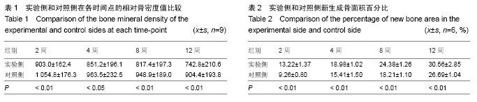
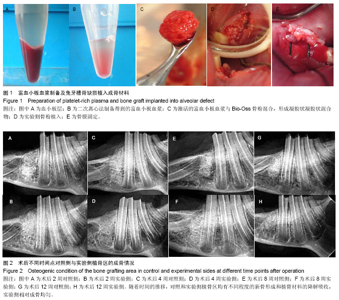
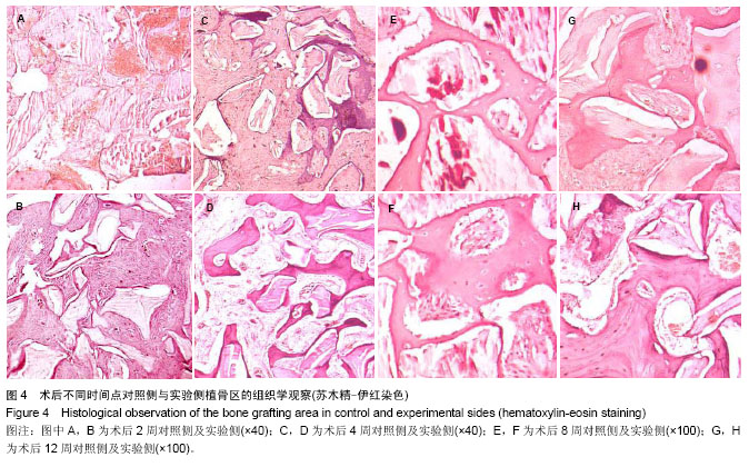
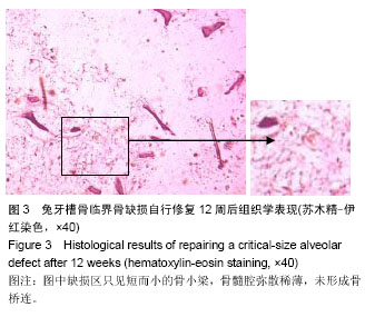
背景:有研究发现,富血小板血浆可促进骨、软组织损伤修复,但在骨愈合过程中的作用仍存在争议,目前尚无定论。
目的:对比观察富血小板血浆复合Bio-Oss/Bio-Gide在兔牙槽骨缺损修复过程中的成骨效果,以探讨富血小板血浆在骨损伤愈合中的作用。
方法:选择新西兰大白兔16只,手术建立兔下颌牙槽骨临界骨缺损模型,采取自身对照,随机选择一侧下颌骨以富血小板血浆/Bio-Oss/Bio-Gide修复骨缺损(实验侧);另外一侧以Bio-Oss/Bio-Gide修复骨缺损(对照侧)。分别于术后2,4,8,12周时各处死4只,通过大体观察、X射线观察、CBCT骨密度测定、组织学切片观察以及新生骨占缺损面积百分比的测定,定性及定量分析牙槽骨缺损区的成骨效果。
结果与结论:术后2-12周的X射线、CBCT及组织学切片观察显示,实验侧和对照侧均有不同程度的新骨形成以及植骨材料的降解吸收;12周时,实验侧缺损表面可见连续的皮质骨形成,植骨区新骨形成并趋于成熟,植骨材料降解吸收明显,对照侧修复效果次之。在术后2,4,8,12周各时间点,实验侧骨密度值均低于对照侧(P < 0.05),实验侧新生骨占面积百分比均大于对照侧(P < 0.05)。说明在兔下颌牙槽骨缺损修复中,富血小板血浆/Bio-Oss/Bio-Gide比Bio-Oss/Bio-Gide具有更好的成骨效果,表明富血小板血浆可促进新骨的形成以及Bio-Oss骨粉的降解吸收。


中国组织工程研究
杂志出版内容重点:组织构建;骨细胞;软骨细胞;细胞培养;成纤维细胞;血管内皮细胞;骨质疏松组织工程


全文链接:

关键词: 组织构建, 骨组织工程, 富血小板血浆, 牙槽骨缺损, 临界骨缺损, 引导骨组织再生, Bio-Oss骨粉, Bio-Gide骨膜,

Abstract:

BACKGROUND: Studies have found that the platelet-rich plasma (PRP) can promote bone and soft tissue injury repair, but its effect on the process of bone healing is still controversial.
OBJECTIVE: To contrastively observe the osteogenesis effect of PRP/Bio-Oss/Bio-Gide in the repair process of alveolar bone defect in rabbits, so as to explore the role of PRP in bone healing.
METHODS: Sixteen New Zealand white rabbits were used to establish animal models of critical-size alveolar bone defect. One side was damaged randomly and repaired by PRP/Bio-Oss/Bio-Gide as experimental side, and the other side repaired by Bio-Oss/Bio-Gide as control side. Four animals were executed at each time-point, postoperative weeks 2, 4, 8, 12. Through general observation, X-ray radiograph, Cone Beam CT assessment, histological examination, the osteogenesis effect in the defect region was qualitatively and quantitatively analyzed.
RESULTS AND CONCLUSION: It could be know from each observation index that as time went on, the experimental and control sides had a different degree of new bone formation and the degradation-absorption of bone graft material. At 12 weeks, continuous cortical bone formation was seen on the surface of the experimental side, new bone formed and tended to be mature, obvious degradation of the bone graft was found, but those in the control side were not as good. At each time-point of 2, 4, 8, 12 weeks, the bone mineral density of the experimental side were lower than that of the control side (P < 0.05), but the percentage of new bone area was 
larger than in the experimental side than the control side (P < 0.05). These findings indicate that the PRP/Bio-Oss/ Bio-Gide has a better osteogenesis effect than the Bio-Oss/Bio-Gide in the repair process of alveolar bone defect in rabbits, and PRP can promote new bone formation and degradation-absorption of Bio-Oss.


中国组织工程研究
杂志出版内容重点:组织构建;骨细胞;软骨细胞;细胞培养;成纤维细胞;血管内皮细胞;骨质疏松组织工程


全文链接:

Key words: alveolar bone loss, guided tissue regeneration, platelet-rich plasma

中图分类号: