| [1] Li YZ, Chen XN, Li YC. Zhongguo Guzhi Shusong Zazhi. 2005; 11(3):342-343. 李毅中,陈献南,李炎川.高龄老人股骨颈骨质疏松性骨折的治疗[J].中国骨质疏松杂志,2005,11(3):342-343.[2] Lin JK, Li YZ, Yu HM, et al. Zhonghua Chuangshang Guke Zazhi. 2011;13(9):885-887. 林金矿,李毅中,俞海明,等.利伐沙班预防老年人髋部骨折术后下肢深静脉血栓形成的研究[J].中华创伤骨科杂志,2011, 13(9): 885-887.[3] Xia WB, He SL, Xu L, et al. Rapidly increasing rates of hip fracture in Beijing, China. J Bone Miner Res. 2011;27: 125-129.[4] Leung F, Blauth M, Bavonratanavech S. Surgery for fragility hip fracture-streamlining the process. Osteoporos Int. 2010; 21(Suppl 4):S519-521.[5] González Della Valle A, Slullitel G, Piccaluga F, et al. The precision and usefulness of preoperative planning for cemented and hybrid primary total hip arthroplasty. J Arthroplasty. 2005;20(1):51-58.[6] Cai SQ, Ren XJ, Yan JX, et al. Chongqing Yike Daxue Xuebao. 2012;37(12):1080-1083. 蔡思清,任晓静,颜建湘,等.年龄对股骨近端几何结构的影响及意义[J].重庆医科大学学报,2012,37(12):1080-1083.[7] Li YZ, Li JL, Lin JK, et al. Zhongguo Guzhi Shusong Zazhi. 2010;16(10):738-741. 李毅中,李建龙,林金矿,等.应用CT扫描观察老年股骨近端皮质骨变化的初步研究[J].中国骨质疏松杂志,2010,16(10):738- 741.[8] Li YZ, Zhuang HF, Lin JK, et al. Comparison of proximal femur geometry among the patients with or without fragile fracture of femoral neck. Osteoporos Int. 2011;22(Suppl 5):S710.[9] Li YZ, Zhuang HF, Lin JK, et al. Zhongguo Guzhi Shusong Zazhi. 2012;18(2):87-89. 李毅中,庄华烽,林金矿,等.年龄对股骨颈骨密度和皮质厚度的影响[J].中国骨质疏松杂志,2012;18(2):87-89.[10] Zhung HF, Li YZ, Lin JK, et al. Zhongguo Guzhi Shusong Zazhi. 2011;17(4):324-327. 庄华烽,李毅中,林金矿,等.脆性股骨颈骨折的股骨近端几何结构分析[J].中国骨质疏松杂志,2011,17(4):324-327.[11] Zhou L, Pei BQ, Lv K, et al. Zhonghua Chuangshang Guke Zazhi. 2007;9(1):58-61. 周力,裴葆青,吕坤,等.股骨颈骨折空心钉内固定手术参数规划与评价系统的研究[J].中华创伤骨科杂志,2007,9(1):58-61.[12] Tang GZ. Zhonghua Chuangshang Guke Zazhi. 2007;9(5): 494-495. 唐国智.空心螺钉固定治疗股骨颈骨折[J].中华创伤骨科杂志, 2007,9(5):494-495.[13] Palm H, Lysén C, Krasheninnikoff M, et al. Intramedullary nailing appears to be superior in pertrochanteric hip fractures with a detached greater trochanter: 311 consecutive patients followed for 1 year. Acta Orthop. 2011;82(2):166-170.[14] Palm H, Jacobsen S, Sonne-Holm S, et al. Integrity of the lateral femoral wall in intertrochanteric hip fractures: an important predictor of a reoperation. J Bone Joint Surg Am. 2007;89(3):470-475.[15] Pajarinen J, Lindahl J, Michelsson O, et al. Pertrochanteric femoral fractures treated with a dynamic hip screw or a proximal femoral nail. A randomised study comparing post-operative rehabilitation. J Bone Joint Surg Br. 2005; 87(1):76-81.[16] Takigami I, Matsumoto K, Ohara A, et al. Treatment of trochanteric fractures with the PFNA (proximal femoral nail antirotation) nail system-report of early results. Bull NYU Hosp Jt Dis. 2008;66(4):276-279.[17] Baumgaertner MR, Curtin SL, Lindskog DM, et al. The value of the tip-apex distance in predicting failure of fixation of peritrochanteric fractures of the hip. J Bone Joint Surg Am. 1995;77(7):1058-1064.[18] Della Valle AG, Padgett DE, Salvati EA. Preoperative planning for primary total hip arthroplasty. J Am Acad Orthop Surg. 2005; 13(7):455-462.[19] Li YZ, Lin JK, Zhang J, et al. Significance of proximal femoral computed tomography scanning in the prediction of femoral prosthesis before total hip arthroplasty. Eur J Orthop Surg Traumatol. 2013;23(1):67-72.[20] Li YZ, Li JL, Lin JK, et al. Zhongguo Zuzhi Gongcheng Yanjiu yu Linchuang Kangfu. 2010;14(30):5535-5538. 李毅中,李建龙,林金矿,等.股骨颈内侧径测量法术前选择股骨假体型号[J].中国组织工程研究与临床康复,2010,14(30): 5535- 5538.[21] Li YZ, Li JL, Lin JK, et al. Zhongguo Zuzhi Gongcheng Yanjiu yu Linchuang Kangfu.2010;14(9):1586-1590. 李毅中,李建龙,林金矿,等.股骨峡部在非骨水泥型全髋关节置换中的作用[J].中国组织工程研究与临床康复,2010,14(9): 1586-1590.[22] Noble PC. Biomechanical advances in total hip replacement. In: Niwa S, Perrem SM, Hattori T, eds. Biomechanics in orthopedics. Tokyo: Springer. 1992.[23] Rubin PJ, Leyvraz PF, Aubaniac JM, et al. The morphology of the proximal femur. A three-dimensional radiographic analysis. J Bone Joint Surg Br. 1992;74(1):28-32.[24] Noble PC, Alexander JW, Lindahl LJ, et al. The anatomic basis of femoral component design. Clin Orthop Relat Res. 1988;(235):148-165.[25] Wang W, Wang Y, Cui J. Zhongguo Linchuang Jiepouxue Zazhi. 2003;21(2):125-128. 汪伟,王岩,崔健.正常股骨近端CT测量及其临床意义[J].中国临床解剖学杂志,2003,21(2):125-128.[26] Li YZ, Li JL, Lin JK et al. Zhongguo Zuzhi Gongcheng Yanjiu yu Linchuang Kangfu. 2012;16(52):9702-9706. 李毅中,李建龙,林金矿,等. 髓腔闪烁指数在全髋关节置换测量中的意义[J].中国组织工程研究与临床康复, 2012,16(52): 9702-9706.[27] Noble PC, Box GG, Kamaric E, et al. The effect of aging on the shape of the proximal femur. Clin Orthop Relat Res. 1995; (316):31-44.[28] LEE CE, Lai YS, Cheng CG. Zhonghua Guanjie Waike Zazhi(Dianziban). 2009;3(4):420-426. 李永恩,赖玉树,郑诚功.近端股骨髓腔几何形状的改变对人工髋关节置换术后存活率的影响[J].中华关节外科杂志(电子版), 2009,3(4):420-426.[29] Kobayashi S, Takaoka K, Saito N, et al. Factors affecting aseptic failure of fixation after primary Charnley total hip arthroplasty. Multivariate survival analysis. J Bone Joint Surg Am. 1997;79(11):1618-1627.[30] Aro HT, Alm JJ, Moritz N, et al. Low BMD affects initial stability and delays stem osseointegration in cementless total hip arthroplasty in women: a 2-year RSA study of 39 patients. Acta Orthop. 2012;83(2):107-114.[31] Joshi AB, Markovic L, Hardinge K, et al. Total hip arthroplasty in ankylosing spondylitis: an analysis of 181 hips. J Arthroplasty. 2002;17(4):427-433.[32] Liu HW, Sun JY, Zhang YK, et al. Zhongguo Linchuang Jiepouxue Zazhi. 2011;29(1):67-71. 刘宏伟,孙俊英,张云坤,等.股骨近段髓腔解剖参数测量与不同类型人工股骨假体的选择[J].中国临床解剖学杂志,2011,29(1): 67-71.[33] Griza S, Ueki MM, Souza DH, et al. Thermally induced strains and total shrinkage of the polymethyl-methacrylate cement in simplified models of total hip arthroplasty. J Mech Behav Biomed Mater. 2012;18C:29-36.[34] van der Veen HC, van Jonbergen HP, Poolman RW, et al. Is there evidence for accelerated polyethylene wear in uncemented compared to cemented acetabular components? A systematic review of the literature. Int Orthop. 2013;37(1):9-14.[35] Lindgren V, Garellick G, Kärrholm J, et al. The type of surgical approach influences the risk of revision in total hip arthroplasty: a study from the Swedish Hip Arthroplasty Register of 90,662 total hipreplacements with 3 different cemented prostheses. Acta Orthop. 2012;83(6):559-565.[36] Hailer NP, Weiss RJ, Stark A, et al. Dual-mobility cups for revision due to instability are associated with a low rate of re-revisions due to dislocation: 228 patients from the Swedish Hip Arthroplasty Register. Acta Orthop. 2012;83(6):566-571.[37] Imamura M, Munro NA, Zhu S, et al. Single mini-incision total hip replacement for the management of arthritic disease of the hip: a systematic review and meta-analysis of randomized controlled trials. J Bone Joint Surg Am. 2012;94(20):1897-1905.[38] Reininga IH, Stevens M, Wagenmakers R, et al. Comparison of gait in patients following a computer-navigated minimally invasive anterior approach and a conventional posterolateral approach for total hip arthroplasty: A randomized controlled trial. J Orthop Res. 2013;31(2):288-294. |

.jpg)
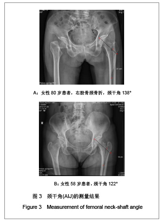
.jpg) 以常用的长度为9 cm的旋转刀刃为例,旋转刀刃尖端将上移0.47 cm,若是股骨近端髋螺钉,则因为螺钉与钢板的夹角为135°,上移将达到1.26 cm,导致尖顶距值增大。因此在做治疗前计划时,应对X射线片的股骨近端结构进行测量,测量颈干角和颈轴长,颈轴长线与股骨头皮质交汇点为顶点(A),选择颈轴长线上距离A点5-10 mm处作为理想旋转刀刃或螺钉尖端点(T),按内固定器械的夹角130°或135°,减去实际测得颈干角得α角,以T为原点,在颈轴长线下方画出与颈轴长线夹角为α的线,此线与股骨外侧皮质交叉点B即为打入旋转刀刃或螺钉点,测量B点与理想尖端点的距离,扣除放大率即为所需旋转刀刃或螺钉长度。这样可以确保尖顶距值在20 mm以内,使螺钉固定在股骨头中心部位,避免穿出或太短,达到理想的内固定效果。
以常用的长度为9 cm的旋转刀刃为例,旋转刀刃尖端将上移0.47 cm,若是股骨近端髋螺钉,则因为螺钉与钢板的夹角为135°,上移将达到1.26 cm,导致尖顶距值增大。因此在做治疗前计划时,应对X射线片的股骨近端结构进行测量,测量颈干角和颈轴长,颈轴长线与股骨头皮质交汇点为顶点(A),选择颈轴长线上距离A点5-10 mm处作为理想旋转刀刃或螺钉尖端点(T),按内固定器械的夹角130°或135°,减去实际测得颈干角得α角,以T为原点,在颈轴长线下方画出与颈轴长线夹角为α的线,此线与股骨外侧皮质交叉点B即为打入旋转刀刃或螺钉点,测量B点与理想尖端点的距离,扣除放大率即为所需旋转刀刃或螺钉长度。这样可以确保尖顶距值在20 mm以内,使螺钉固定在股骨头中心部位,避免穿出或太短,达到理想的内固定效果。