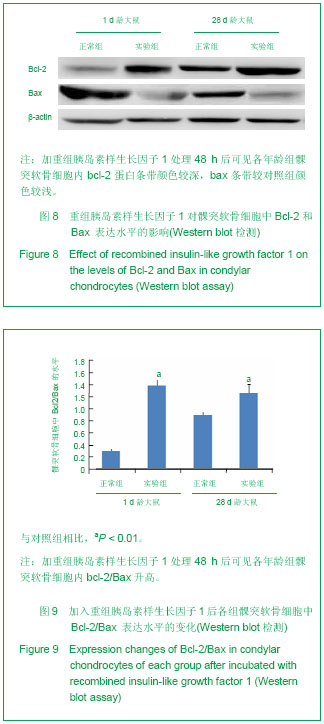
| [1] Singh M, Detamore MS. Biomechanical properties of the mandibular condylar cartilage and their relevance to the TMJ disc. J Biomech. 2009;42(4):405-417. [2] Kuroda S, Tanimoto K, Izawa T, et al. Biomechanical and biochemical characteristics of the mandibular condylar cartilage. Osteoarthritis Cartilage. 2009;17(11):1408-1415.[3] Giustina A, Mazziotti G, Canalis E. Growth hormone, insulin-like growth factors, and the skeleton. Endocr Rev. 2008;29(5):535-559. [4] Ohlsson C, Mohan S, Sjögren K, et al. The role of liver-derived insulin-like growth factor-I. Endocr Rev. 2009;30(5): 494-535. [5] Maor G, Hochberg Z, Silbermann M. Insulin-like growth factor I accelerates proliferation and differentiation of cartilage progenitor cells in cultures of neonatal mandibular condyles. Acta Endocrinol (Copenh). 1993;128(1):56-64.[6] Visnapuu V, Peltomäki T, Rönning O, et al. Distribution of insulin-like growth factor-I mRNA in the mandibular condyle and rib cartilage of the rat during growth. Arch Oral Biol. 2002; 47(11):791-798.[7] Ohlsson C, Sjögren K, Jansson JO, et al. The relative importance of endocrine versus autocrine/paracrine insulin-like growth factor-I in the regulation of body growth. Pediatr Nephrol. 2000;14(7):541-543.[8] Datta HK, Ng WF, Walker JA, et al. The cell biology of bone metabolism. J Clin Pathol. 2008;61(5):577-587. [9] Shibata S, Fukuoka H, Sato R, et al. An in situ hybridization study of the insulin-like growth factor system in developing condylar cartilage of the fetal mouse mandible. Eur J Histochem. 2012;56(2):e23. [10] Hinton RJ, Serrano M, So S. Differential gene expression in the perichondrium and cartilage of the neonatal mouse temporomandibular joint. Orthod Craniofac Res. 2009;12(3): 168-177. [11] Sriram D, Jones A, Alatli-Burt I, et al. Effects of mechanical stimuli on adaptive remodeling of condylar cartilage. J Dent Res. 2009;88(5):466-470.[12] Fuentes MA, Opperman LA, Bellinger LL, et al. Regulation of cell proliferation in rat mandibular condylar cartilage in explant culture by insulin-like growth factor-1 and fibroblast growth factor-2. Arch Oral Biol. 2002;47(9):643-654.[13] Higgins TF, Johnson BD. Effect of exogenous IGF-1 on chondrocyte apoptosis in a rabbit intraarticular osteotomy model. J Orthop Res. 2010;28(1):125-130. [14] Hajjar D, Santos MF, Kimura ET. Propulsive appliance stimulates the synthesis of insulin-like growth factors I and II in the mandibular condylar cartilage of young rats. Arch Oral Biol. 2003;48(9):635-642.[15] Chen Y, Ke J, Long X, et al. Insulin-like growth factor-1 boosts the developing process of condylar hyperplasia by stimulating chondrocytes proliferation. Osteoarthritis Cartilage. 2012; 20(4): 279-287.[16] Yokota T, Shimokawa H, Shibata S, et al. Insulin-like growth factor I regulates apoptosis in condylar cartilage. J Dent Res. 2008;87(2):159-163.[17] Götz W, Kunert D, Zhang D, et al. Insulin-like growth factor system components in the periodontium during tooth root resorption and early repair processes in the rat. Eur J Oral Sci. 2006;114(4):318-327.[18] Matsuda S, Mishima K, Yoshimura Y, et al. Apoptosis in the development of the temporomandibular joint. Anat Embryol (Berl). 1997;196(5):383-391.[19] Watahiki J, Yamaguchi T, Irie T, et al. Gene expression profiling of mouse condylar cartilage during mastication by means of laser microdissection and cDNA array. J Dent Res. 2004;83(3):245-249.[20] 江莉婷,朱雅萍,魏立,等.大鼠髁突软骨细胞冻存复苏后的生物学特性观察[J].现代口腔医学杂志,2012,26(4):251-257。[21] Livak KJ, Schmittgen TD. Analysis of relative gene expression data using real-time quantitative PCR and the 2(-Delta Delta C(T)) Method. Methods. 2001;25(4):402-408.[22] Meikle MC. Remodeling the dentofacial skeleton: the biological basis of orthodontics and dentofacial orthopedics. J Dent Res. 2007;86(1):12-24.[23] Sriram D, Jones A, Alatli-Burt I, et al. Effects of mechanical stimuli on adaptive remodeling of condylar cartilage. J Dent Res. 2009;88(5):466-470.[24] Loreto C, Almeida LE, Trevilatto P, et al. Apoptosis in displaced temporomandibular joint disc with and without reduction: an immunohistochemical study. J Oral Pathol Med. 2011;40(1):103-110.[25] Ohshima T, Yonezu H, Nishibori Y, et al. Morphological observation of process of mouse temporomandibular joint formation. Bull Tokyo Dent Coll. 2011;52(4):183-190.[26] Caltabiano R, Leonardi R, Musumeci G, et al. Apoptosis in temporomandibular joint disc with internal derangement involves mitochondrial-dependent pathways. An in vivo study. Acta Odontol Scand. 2013;71(3-4):577-583. [27] Yokota T, Shimokawa H, Shibata S, et al. Insulin-like growth factor I regulates apoptosis in condylar cartilage. J Dent Res. 2008;87(2):159-163.[28] Mackey TJ, Borkowski A, Amin P, et al. bcl-2/bax ratio as a predictive marker for therapeutic response to radiotherapy in patients with prostate cancer. Urology. 1998;52(6):1085-1090.[29] Petros AM, Olejniczak ET, Fesik SW. Structural biology of the Bcl-2 family of proteins. Biochim Biophys Acta. 2004;1644 (2-3): 83-94.[30] Wu MJ, Zhan J, Gu ZY. Time course of expression of bcl-2 and bax in rabbit condylar chondrocytes following forward mandibular positioning. Angle Orthod. 2008;78(3):453-459. [31] Wu M, Gu Z, Xiao J, et al. Differential expression of apoptosis-associated proteins in chondrocytes of the mandibular condyles of rabbits with anterior disk displacement. Cranio. 2008;26(2):144-149.[32] Huang Q, Singh B, Sharawy M. Immunohistochemical analysis of Bcl-2 and Bax oncoproteins in rabbit craniomandibular joint. Arch Oral Biol. 2004;49(2):143-148.[33] Trump BF, Berezesky IK, Chang SH, et al. The pathways of cell death: oncosis, apoptosis, and necrosis. Toxicol Pathol. 1997;25(1):82-88.[34] Elmore S. Apoptosis: a review of programmed cell death. Toxicol Pathol. 2007;35(4):495-516.[35] Loreto C, Almeida LE, Migliore MR, et al. TRAIL, DR5 and caspase 3-dependent apoptosis in vessels of diseased human temporomandibular joint disc. An immunohistochemical study. Eur J Histochem. 2010;54(3):e40.[36] Loreto C, Musumeci G, Leonardi R. Chondrocyte-like apoptosis in temporomandibular joint disc internal derangement as a repair-limiting mechanism. An in vivo study. Histol Histopathol. 2009;24(3):293-298.[37] Ciarmatori S, Kiepe D, Haarmann A, et al. Signaling mechanisms leading to regulation of proliferation and differentiation of the mesenchymal chondrogenic cell line RCJ3.1C5.18 in response to IGF-I. J Mol Endocrinol. 2007; 38(4):493-508.[38] Navarro M, Baserga R. Limited redundancy of survival signals from the type 1 insulin-like growth factor receptor. Endocrinology. 2001;142(3):1073-1081.[39] Qiao M, Shapiro P, Kumar R, et al. Insulin-like growth factor-1 regulates endogenous RUNX2 activity in endothelial cells through a phosphatidylinositol 3-kinase/ERK-dependent and Akt-independent signaling pathway. J Biol Chem. 2004;279 (41): 42709-42718. [40] Prasadam I, Mao X, Wang Y, et al. Inhibition of p38 pathway leads to OA-like changes in a rat animal model. Rheumatology (Oxford). 2012;51(5):813-823. [41] Bobick BE, Kulyk WM. Regulation of cartilage formation and maturation by mitogen-activated protein kinase signaling. Birth Defects Res C Embryo Today. 2008;84(2):131-154. |
.jpg)
.jpg)