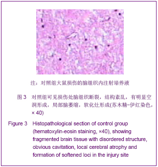
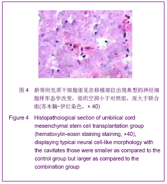
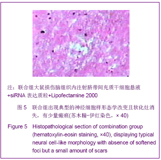
| [1] Arabi YM, Haddad S, Tamim HM,et al. Mortality reduction after implementing a clinical practice guidelines–based management protocol for severe traumatic brain injury. J Crit Care.2010;25(2):190-195.[2] Lee HC, Chuang HC, Cho DY, et al. Applying Cerebral Hypothermia and Brain Oxygen Monitoring in Treating Severe Traumatic Brain Injury. World Neurosurg. 2010;74(6): 654-660.[3] Engelmann CM, Siert L. Cognitive disturbances following severe traumatic brain injury. Ugeskr Laege. 2007; 169(3): 217-219.[4] Liliang PC, Liang CL, Weng HC, et al. Proteins in Serum Predict Outcome After Severe Traumatic Brain Injury. J Surg Res. 2010;160(2): 302-3078.[5] Sekhon MS, Dhingra VK, Sekhon IS,et al.The safety of synthetic colloid in critically ill patients with severe traumatic brain injuries. J Crit Care.2011;26(4):357-362.[6] Yoo BY, Shin YH, Yoon HH,et al.Application of mesenchymal stem cells derived from bone marrow and umbilical cord in human hair multiplication.J Dermatol Sci. 2010, 60(2): 74-83.[7] Xu Y,Meng H,Li C,et alUmbilical cord-derived mesenchymal stem cells isolated by a novel explantation technique can differentiate into functional endothelial cells and promote revascularization. Stem Cells.2010;19(10):1511-1522. [8] Matsuse D,Kitada M,Kohama M,et al. Human umbilical cord-derived mesenchymal stromal cells differentiate into functional Schwann cells that sustain peripheral nerve regeneration.J Neuropathol Exp Neurol.2010;69(9):973-985.[9] Yuan GF, Li HX. Zhongguo Xiandai Yixue Zazhi. 2011;21(13): 1435-1438.苑国富,李红星.大鼠颅脑损伤后亚低温治疗对RhoA 及Nogo- A 表达的影响[J].中国现代医学杂志,2011, 21(13):1435-1438.[10] Wang QY, Liu WG, Wang ZY. Zhongguo Guke Linchuang yu Jichu Yanjiu Zazhi.2010;2(4):292-296.王求永,刘文革,王振宇.FTY720对大鼠急性脊髓损伤后RhoA 表达的影响[J].中国骨科临床与基础研究杂志,2010,2(4):292-296.[11] Ahmed Z, Dent RG, Suggate EL, et al. Disinhibition of neurotrophin-induced dorsal root ganglion cell neurite outgrowth on CNS myelin by siRNA-mediated knockdown of NgR, p75NTR and Rho-A. Molecular and Cellular Neuroscience.2005;28(3):509-523.[12] Chen L,Hui GZ,Zhang S,et al.Zhonghua Chuangshang Zazhi. 2009;25(6):498-502.陈镭,惠国桢,张赛,等.人脐血间充质干细胞移植促进大鼠颅脑损伤后神经功能的恢复[J].中华创伤杂志,2009,25(6):498- 502.[13] Horisawa T, Ishibashi T, Nishikawa H, et al. The effects of selective antagonists of serotonin 5-HT7 and 5-HT1A receptors on MK-801-induced impairment of learning and memory in the passive avoidance and Morris water maze tests in rats: Mechanistic implications for the beneficial effects of the novel atypical antipsychotic lurasidone . Behavioural Brain Res. 2011;220 (1): 83-90.[14] Mutlu O, Ulak G, Celikyurt IK et al, Effects of olanzapine, sertindole and clozapine on learning and memory in the Morris water maze test in naive and MK-801-treated mice.Pharmaco Biochem Behav. 2011;98(3): 398-404.[15] Li L, Ding J, Marshall C, et al.Pretraining affects Morris water maze performance with different patterns between control and ovariectomized plus d-galactose-injected mice. Behavioural Brain Research.2011;217(1): 244-247.[16] Hang RH, Xu YJ, Xie HF.Evaluating on recognition impairment after traumatic brain injury with WCST.Fa Yi Xue Za Zhi. 2011;27(5):346-349. [17] Han SR, Yee GT, Choi CY,et al.Cortical laminar necrosis in an infant with severe traumatic brain injury.J Korean Neurosurg Soc. 2011;50(5):472-474. [18] Shultz SR, Macfabe DF, Foley KA, et al.Sub-concussive brain injury in the Long-Evans rat induces acute neuroinflammation in the absence of behavioral impairments.Behav Brain Res. 2011;229(1):145-152. [19] Settervall CH, Sousa RM, Silva SC.In-hospital mortality and the Glasgow Coma Scale in the first 72 hours after traumatic brain injury.Rev Lat Am Enfermagem. 2011;19(6):1337-1343. [20] Yamashita S, Hasuo H, Tokutomi T,et al. Edaravone attenuates impairment of synaptic plasticity in granule cell layer of the dentate gyrus following traumatic brain injury. Kurume Med J. 2011;58(2):47-58.[21] Han SR, Yee GT, Choi CY, et al Cortical laminar necrosis in an infant with severe traumatic brain injury.J Korean Neurosurg Soc. 2011;50(5):472-474. [22] Hang RH, Xu YJ, Xie HF,et al. Evaluating on recognition impairment after traumatic brain injury with WCST.Fa Yi Xue Za Zhi. 2011;7(5):346-349. [23] He JT, Mang J, Mei CL,et al. Neuroprotective effects of exogenous activin a on oxygen-glucose deprivation in PC12 cells.Molecules. 2011;30(17):315-327.[24] Li GX,Wang CM,Gong LP,et al.Zhongguo Linchuang Shenjing Waike Zazhi. 2009,14(5):288-290.黎国雄,王传湄, 龚利平,等.神经干细胞立体定向脑内移植治疗大鼠重型颅脑损伤[J].中国临床神经外科杂志,2009,14(5): 288-290.[25] Zhao EY, Wang LD, Wen QQ, et al. Fasudil hydrochloridedifferentiates bone marrow mesenchymal stem cells into neurons via Notch signaling. Neural Regen Res. 2010;5(11):814-819.[26] Dergham P,Ellezam B,Essagian C,et al.Rho signaling pathwaytargeted to promote spinal cord repair. J Neurosci. 2002;22(15):6570-6577. [27] Govek EE,Newey SE,Aelstl V. The role of the Rho GTPases in neuronal development. Genes Dev.2005;19(10): 1-49.[28] Neuhof S,Moers J,Ricks M. Proliferation, diferentiation, and cytokine secretion of human umbilical cord blood-derivedmononuclear ceils in vitro.Exp Hematol.2007; 35(7):1119-1131.[29] Pang KM, Sung MA, Alrash-dan MS, et al. Trans-plantation of mesenchymal stem cells from human umbilical cord versus human umbilical cord blood for peripheral nerve regenera-tion. Neural Regen Res. 2010;5(11):838-845.[30] Ha Y,Choi JU,Yoon DH,et a1.Neural phenotype expression of cultured human cord blood cells in vitro. Neurureport. 2001; 12(16):3523-3530.[31] Buzahska L,Jurga M,Domafiska-Janik K.Neumnal differentiation of human umbilical cord blood neural stem- like cell line.Neurodegener Dis.2006;3(1-2):19-26.[32] Wang D,Zhang JJ,Mang JJ.Zhongguo Zuzhi Gongcheng Yanjiu yu Linchuang Kangfu. 2010,14(14): 2539-2544.王东,张建军,马景鑑.神经干细胞NgR基因沉默立体定向移植治疗大鼠脑损伤[J].中国组织工程研究与临床康复,2010, 14(14): 2539-2544.[33] Neuhof S,Moers J,Ricks M.Proliferation,diferentiation,and cytokine secretion of human umbilical cord blood-derived mononuclear ceils in vitro.Exp Hematol.2007;35(7): 1119-1131.[34] Zhou GQ,Jin Y,Zhang P.Zhongguo Zuzhi Gongcheng Yanjiu yu Linchuang Kangfu. 2010;14(45):8416-8420.周国庆,金怡,张鹏.沉默RhoA基因对骨髓间充质干细胞静脉移植治疗脑梗死大鼠的作用[J].中国组织工程研究与临床康复, 2010,14(45):8416-8420. |