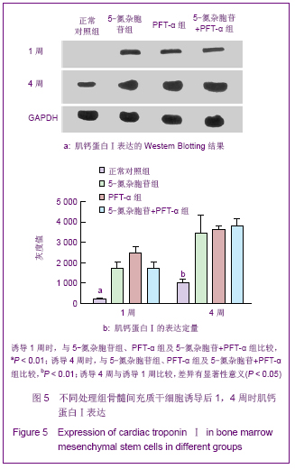
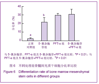
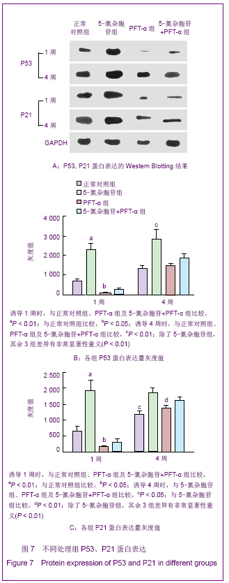
| [1] Elizabeth GN. Cardiovascular disease. New England J Med. 2003;349:60-72.[2] Cao F, Sun DD, Li CX, et al. Long-term myocardial functional improvement after autologous bone marrow mononuclear cells transplantation in patients with ST-segment elevation myocardial infarction: 4 years follow-up. Eur Heart J. 2009; 30(16):1986-1994.[3] Deng W, Obrocka M, Fischer I, et al. In vitro differentiation of human marrow stromal cells into early progenitors of neural cells by conditions that increase intracellular cyclic AMP. Biochem Biophys Res Commun. 2001;282(1): 148-152.[4] Wakitani S, Saito T, Caplan AI. Myogenic cells derived from rat bone marrow mesenchymal stem cells exposed to 5-azacytidine. Muscle Nerve. 1995;18:1417-1426.[5] Makino S, Fukuda K, Miyoshi S, et al. Cardiomyocytes can be generated from marrow stromal cells in vitro. J Clin Invest. 1999;103(5):697-705. [6] Hong H, Takahashi K, Ichisaka T, et al. Suppression of induced pluripotent stem cell generation by the p53-p21 pathway. Nature. 2009;460: 1132-1136.[7] Armesilla DA, Elvira G, Silva A. p53 regulates the proliferation, differentiation and spontaneous transformation of mesenchymal stem cells. Exp Cell Res. 2009;315: 3598-3610.[8] Pulukuri SM, Rao JS. Activation of p53/p21Waf1/Cip1 pathway by 5-aza-2'-deoxycytidine inhibits cell proliferation, induces pro-apoptotic genes and mitogen-activated protein kinases in human prostate cancer cells. Int J Oncol. 2005;26(4): 863-871.[9] Zhu WG, Hileman T, Ke Y, et al. 5-Aza-2,-deoxycytidine Activates the p53/p21Waf1/Cip1 Pathway to Inhibit Cell Proliferation. J Biol Chem. 2004;279(15): 15161-15166.[10] The Ministry of Science and Technology of the People’s Republic of China. Guidance Suggestions for the Care and Use of Laboratory Animals. 2006-09-30.[11] Bergmann O, Bhardwaj RD, Bernard S, et al. Evidence for Cardiomyocyte Renewal in Humans. Science. 2009; 324 (5923): 98 -102.[12] Li X,Yu X, Lin Q, et al. Bone marrow mesenchymal stem cells differentiate into functional cardiac phenotypes by cardiac microenvironment. J Mol Cell Cardiol. 2007;42(2):295-303. [13] Wang JS, Shunr Tim D, Galipeeall J, et al. Marrou Stramol cells for cellular Cardiomyoplasty :feasibillty and potential dinical advantages. Thorae Cardiovasc Surg. 2000;120(62): 999-1005.[14] Strauer BE, Brehm M, Zeus T, et al. Repair of infarcted myocardium by autologous intracoronary mononuclear bone marrow cell transplantation in humans. Circulation. 2002;106 (15):1913-1918.[15] Janssens S, Dubois C, Bogaert J, et al. Autologous bone marrow-derived stem-cell transfer in patients with ST-segment elevation myocardial infarction:double-blind, randomised controlled trial. Lancet. 2006;367(9505): 113-121. [16] Wollert KC, Meyer GP, Lotz J, et al. Intracoronary autologous bone marrow cell transfer after myocardial infarction: The BOOST randomised controlled clinical trial. Lancet. 2004; 364(9429) :141-148.[17] Lago N, Trainini J, Christen A, et al. Mononuclear bone marrow stem cells implant as an alternative treatment in non-ischemic dilated cardiomyopathy. J Am Coll Cardiol. 2006;47(4 Suppl. A):1A-384A.[18] Wang J, Xie X, He H, et al. A prospective, randomized, controlled trial of autologous mesenchymal stem cells transplantation for dilated cardiomyopathy. Chin J Cardiol. 2006;34(2):107-111. [19] Komarova EA, Gudkov AV. Suppression of p53: a new app roach to overcome side effects of antitumor therapy. Biochemistry (Mosc).2000;65:41-48.[20] Schneider SR, Diab AM, Rohrbeck A, et al. 5-aza-Cytidine is a potent inhibitor of DNA Methyltransferase 3a and induces apoptosis in HCT-116 colon cancer cells via Gadd45- and p53-Dependent Mechanisms. J Pharmacol Exp Ther. 2005; 312(2):525-536.[21] Yan XB, Lü AL, Liu BW, et al.Zhongguo Zuzhi Gongcheng Yanjiu yu Linchuang Kangfu. 2011;15(14):2482-2486.燕学波,吕安林,刘博武,等. P53-P21蛋白通路对5-氮杂胞苷诱导的大鼠骨髓间充质干细胞增殖和凋亡的影响[J].中国组织工程研究与临床康复,2011,15(14):2482-2486.[22] Levine AJ, Finlay CA, Hinds PW. P53 is a Tumor Suppressor Gene. Cell. 2004;116:67-69.[23] Schuler M, Green DR. Transcription, apoptosis and P53: catch-22. Trends Genet. 2005;21(3): 182-187.[24] Xuwan L, Chu CC, Jinping G, et al. Pifithrin-α protects against doxorubicin-induced apoptosis and acute cardiotoxicity in mice. Am J Physiol Heart Circ Physiol. 2004;286: 933-939.[25] Shave R, Baggish A, George K, et al. Exercise-induced cardiac troponin elevation: evidence, mechanisms, and implications. J Am Coll Cardiol. 2010;56(3):169-176. [26] Janczarska K, Kie?-Wilk B, Leszczyńska-Go?abek I, et al. The role of connexin 43 in preconditioning. Impact on mitochondrial function. Kardiol Pol. 2010;68(1):91-96. |
.jpg)