2.1 兔骨髓间充质干细胞的分离培养、鉴定及诱导的细胞形态变化 骨髓间充质干细胞接种时细胞呈悬浮的球形细胞,小圆形。接种24 h后即可观察到个别细饱贴壁,大多呈圆形或椭圆形。细胞培养至14 d时接近融合,胞融合成细胞排列有一定的方向性,呈漩涡状排列,呈成纤维细胞样。细胞贴壁达80%时开始传代,经传代的兔骨髓间充质干细胞,3次传代后细胞形态稳定,呈长梭形,为成纤维细胞样,与原代细胞无差别。见图1。
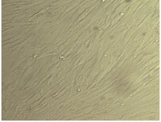
Figure 1 Passage 3 bone marrow mesenchymal stem cells exhibit long spindle-shaped and fibroblast-like appearance (×200)
图1 兔骨髓间充质干细胞3次传代后细胞形态稳定,呈长梭形,为成纤维细胞样(×200)
经过流式细胞仪检测结果为细胞表面抗原CD29、CD44阳性细胞数分别占99.54%和97.87%,结果证实了培养的细胞符合兔骨髓间充质干细胞表面标志特点。见图2。
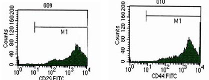
Figure 2 Expression of CD antigen on the surface of bone marrow mesenchymal stem cells with flow cytometry analysis
图2 流式细胞仪检测兔骨髓间充质干细胞表面CD抗原表达情况
骨髓间充质干细胞在诱导分化第7天左右,光镜下可以发现胞体变大,由长梭形逐渐变为短梭形,且细胞出现粗颗粒。随着诱导时间的延长,至21 d时发现细胞由短梭形向类圆形、圆形细胞转变,胞浆丰富。见图3。
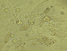
Figure 3 The bone marrow mesenchymal stem cells showed similar round shape after induced for 21 d (×400)
图3 诱导至21 d时,兔骨髓间充质干细胞呈类圆形细胞(×400)
2.2 诱导后兔骨髓间充质干细胞的形态学变化及功能鉴定结果 添加肝细胞生长因子和表皮生长因子诱导因子后于14 d时行甲胎蛋白染色,表现为胞浆染成红棕色。见图4。
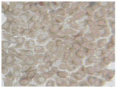
Figure 4 Alpha-fetoprotein staining of bone marrow mesenchymal stem cells showed brown-red color after induced for 21 d (×200)
图4 诱导至14 d时兔骨髓间充质干细胞甲胎蛋白染色呈棕红色(×200)
血清白蛋白染色,糖原染色在诱导后第21天时发现呈阳性,分别表现为胞浆内呈红棕色和紫红色。见图5,6。
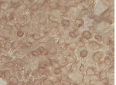
Figure 5 Bone marrow mesenchymal stem cells showed brown-red color by serum albumin staining after induced for 21 d (×200)
图5 诱导至21 d时兔骨髓间充质干细胞血清白蛋白染色为棕红色(×200)
血清白蛋白染色,糖原染色在诱导后第21天时发现呈阳性,分别表现为胞浆内呈红棕色和紫红色。见图5,6。
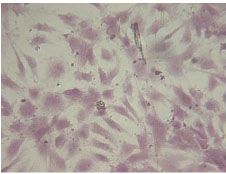
Figure 6 Glycogen staining of the bone marrow mesenchymal stem cells after induced for 21 d (×200)
图6 诱导至21 d时兔骨髓间充质干细胞糖原染色效果(×200)
2.3 肝硬化模型的建立结果 28只普通级兔在造模过程中最终死亡5只,死亡率为17.86%。于实验8周末随机处死2只。正常兔大体上观察,肝脏质地柔软光滑;8周末可见肝脏体积缩小,表面呈小结节或小颗粒状。见图7,8。
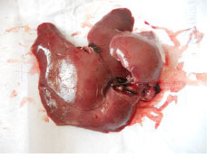
Figure 7 Normal liver specimens , 图7 正常兔肝脏标本
2.3 肝硬化模型的建立结果 28只普通级兔在造模过程中最终死亡5只,死亡率为17.86%。于实验8周末随机处死2只。正常兔大体上观察,肝脏质地柔软光滑;8周末可见肝脏体积缩小,表面呈小结节或小颗粒状。见图7,8。
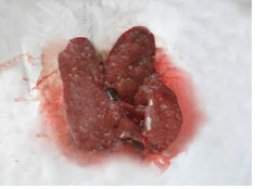
Figure 8 Specimens of rabbit liver cirrhosis
图8 肝硬化兔肝脏标本
正常兔肝脏肝小叶由肝细胞板围绕中央静脉呈放射状排列,肝板间为肝窦填充。见图9。

Figure 9 Typical hepatic lobule could be seen in the normal liver tissue (×40)
图9 正常兔肝组织,可见典型的肝小叶(×40)
将诱导后BrdU标记的肝样细胞经门静脉内移植结果 以门静脉内移植的方式移植的2×10
6个诱导后经BrdU标记的肝样细胞见图9,10。移植2 d时,实验组死亡1只。
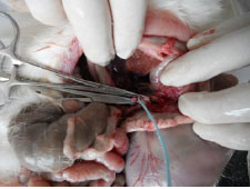
Figure 10 Induced hepatocyte-like cells were injected into the bone marrow mesenchymal stem cells through portal vein
图10 诱导后的肝样细胞经门静脉内注入
BrdU免疫组织化学染色显示:在汇管区和肝索肝窦内均可见BrdU阳性细胞,染色细胞呈棕黑色。肝脏病理显示:汇管区周围有再生细胞团。见图11,12。
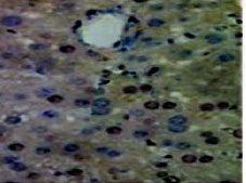
Figure 11 BrdU labeled hepatocyte-like cells showed brown-black color after injected through portal vein as shown by immunohistochemical staining (×200)
图11 BrdU标记肝样细胞经门静脉后经免疫组织化学染色为棕黑色(×200)
BrdU免疫组织化学染色显示:在汇管区和肝索肝窦内均可见BrdU阳性细胞,染色细胞呈棕黑色。肝脏病理显示:汇管区周围有再生细胞团。见图11,12
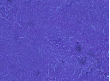
Figure 12 Image of the tissue section at 21 d after transplantation, newborn liver cells can be seen between the pseudolobules (×40)
图12 细胞移植21 d组织切片,在假小叶之间可以看到新生肝细胞(×40)
长期皮下注射CCl
4造成肝硬化。移植第4-21天,与对照组比较,
P < 0.05,在移植细胞14 d时已接近正常范围(建模前随机选10只兔抽取血验肝功所得):(43.8±12.3) U/L,(52.5±10.3) U/L。细胞移植从第14天时开始血清白蛋白达(34.6±12.9) g/L,与对照组比较差异有显著性意义(
P < 0.05),随后血清白蛋白在正常范围(34.2±9.2) g/L,总蛋白与血清白蛋白变化情况一致。见表1。
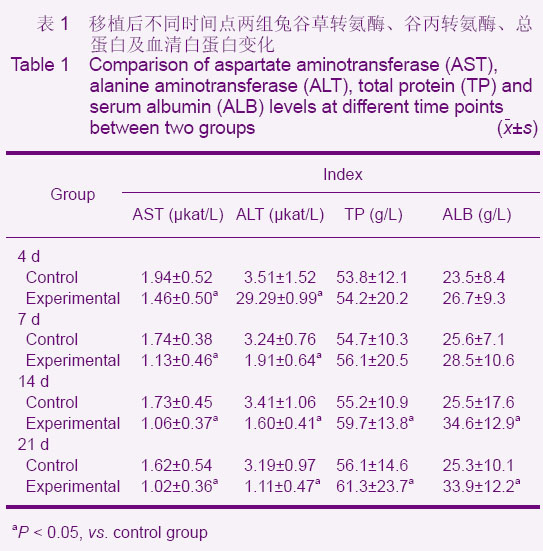
表1 移植后不同时间点两组兔谷草转氨酶、谷丙转氨酶、总蛋白及血清白蛋白变化
Table 1 Comparison of aspartate aminotransferase (AST), alanine aminotransferase (ALT), total protein (TP) and serum albumin (ALB) levels at different time points between two groups (x±s)
