中国组织工程研究 ›› 2019, Vol. 23 ›› Issue (36): 5850-5855.doi: 10.3969/j.issn.2095-4344.1944

• 骨与关节图像与影像 bone and joint imaging • 上一篇    下一篇

双侧椎弓根螺钉置入内固定联合椎间融合治疗腰椎间盘突出症:CT影像学的客观评估

郑淞文1,孟  仪2,龙钰函1,徐  哲1,张泳华1   

  1. 大连市中心医院,1放射科,2脊柱外科,辽宁省大连市  116033
  • 出版日期:2019-12-28 发布日期:2019-12-28
  • 作者简介:郑淞文,女,1980年生,山东省荣成市人,汉族,2011年大连医科大学毕业,硕士,副主任医师,主要从事神经与骨肌影像诊断方面的研究。

Bilateral pedicle screw fixation combined with interbody fusion for lumbar disc herniation: objective evaluation of CT imaging

Zheng Songwen1, Meng Yi2, Long Yuhan1, Xu Zhe1, Zhang Yonghua1
  

  1. 1Department of Radiology, 2Department of Spine Surgery, Dalian Central Hospital, Dalian 116033, Liaoning Province, China
  • Online:2019-12-28 Published:2019-12-28
  • About author:Zheng Songwen, Master, Associate chief physician, Department of Radiology, Dalian Central Hospital, Dalian 116033, Liaoning Province, China

摘要:

文章快速阅读:


文题释义:
腰椎间盘突出:是较为常见的疾患之一,主要是因为腰椎间盘各部分(髓核、纤维环及软骨板),尤其是髓核,有不同程度的退行性改变后,在外力因素的作用下,椎间盘的纤维环破裂,髓核组织从破裂之处突出(或脱出)于后方或椎管内,导致相邻脊神经根遭受刺激或压迫,从而产生腰部疼痛,一侧下肢或双下肢麻木、疼痛等一系列临床症状。腰椎间盘突出症以L4-5、L5-S1发病率最高,约占95%。
双侧椎弓根螺钉固定融合:被公认为处理各类脊柱疾病的标准方案,其具有生物力学及临床优势,双侧椎板关节突螺钉固定联合椎间融合器置入具有与双侧椎弓根螺钉固定联合椎间融合器置入相同的力学稳定性,增加脊柱的稳定性。
 
摘要
背景:对于合并有显著的腰椎不稳定的患者,需要通过外科手术将突出的椎间盘部分切除,同时扩大变窄的神经根管以缓解腰神经根的压迫,并且在切除突出椎间盘和扩大神经根管的同时还需要内固定联合植骨融合以恢复脊柱的稳定性,以解除不稳定因素所导致的腰神经根受压迫而产生的腰腿疼痛症状。有研究认为,CT检查可以准确的反映椎间盘是否发生突出,清楚显示椎间盘突出的形态、大小和部位。
目的:CT影像学评估双侧椎弓根螺钉置入内固定联合椎间融合治疗腰椎间盘突出症的临床疗效。
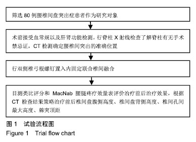
方法:回顾性分析2015年8月至2017年7月在大连市中心医院接受治疗的80例腰椎间盘突出症患者的病历资料。所有患者均接受双侧椎弓根螺钉置入内固定联合椎间融合治疗。研究于2018-06-30通过大连市中心医院伦理委员会审核批准。
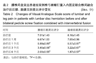
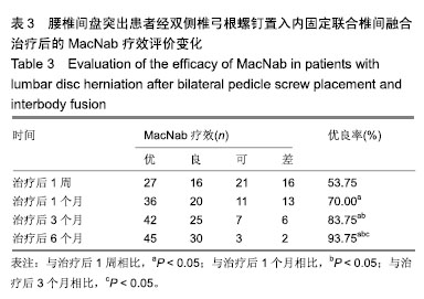
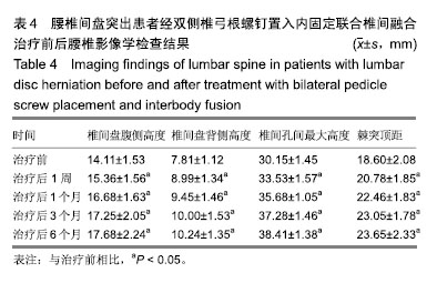
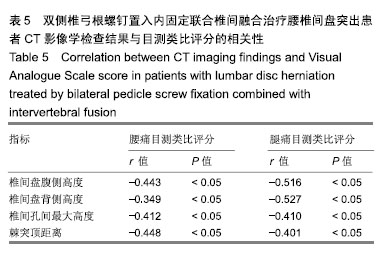
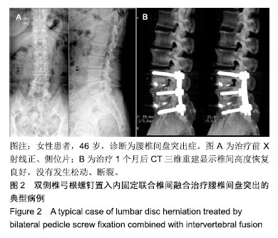
结果与结论:①所有患者均顺利完成手术且完成随访;②治疗后1,3个月和末次随访的腰腿痛目测类比评分均显著低于治疗前(P < 0.05);③随着时间的延长,腰椎间盘突出患者治疗后MacNab疗效的优良率逐渐显著增加(P < 0.05);④治疗后各时间点的椎间盘腹侧高度、椎间盘背侧高度、椎间孔间最大高度、棘突顶距均显著性高于治疗前(P < 0.05);影像学显示治疗后患者的腰椎角度逐渐恢复,病变节段椎间隙高度恢复,CT扫描检查显示椎弓根螺钉固定良好,没有断裂和松动现象;治疗前及治疗后随访,CT影像检查的椎间盘腹侧高度、椎间盘背侧高度、椎间孔间最大高度、棘突顶距离与腰痛目测类比评分、腿痛目测类比评分之间均呈负相关(P < 0.05);⑤结果提示采用双侧椎弓根螺钉置入内固定联合椎体融合治疗腰椎间盘突出的临床疗效显著,使用CT影像学方法不但可以评估病情而且还可以评估椎间盘腹侧高度、椎间盘背侧高度、椎间孔间最大高度、棘突顶距恢复情况、腰椎角度恢复情况、螺钉置入的位置以及鉴别螺钉是否发生松动或断裂,评估临床疗效。


ORCID: 0000-0002-3450-3269(郑淞文)

关键词: 腰椎间盘突出症, 双侧椎弓根螺钉置入内固定, 椎间融合, CT, 椎间盘腹侧高度, 椎间盘背侧高度, 椎间孔间最大高度, 棘突顶距

Abstract:

BACKGROUND: For patients with significant lumbar instability, surgical resection of the herniated intervertebral disc and enlargement of the narrowed nerve root canal are needed to relieve the compression of the lumbar nerve root. Simultaneously, internal fixation and bone graft fusion are needed to restore the stability of the spine when resecting the herniated intervertebral disc and enlarging the nerve root canal so as to relieve the symptoms of lumbar and leg pain caused by compression of lumbar nerve root caused by instability factors. Some studies believe that CT examination can accurately reflect whether the intervertebral disc protrusion occurs, and clearly show the shape, size and location of the intervertebral disc protrusion.

OBJECTIVE: CT imaging was used to evaluate clinical efficacy of bilateral pedicle screw fixation combined with interbody fusion for lumbar disc herniation.
METHODS: Data of 80 cases of lumbar disc herniation treated in Dalian Municipal Central Hospital from August 2015 to July 2017 were retrospectively analyzed. All patients received bilateral pedicle screw fixation combined with interbody fusion. This study was approved by the Ethics Committee of Dalian Central Hospital on June 30, 2018.
RESULTS AND CONCLUSION: (1) The operation was successfully performed in all patients. All patients were followed up. (2) The Visual Analogue Scale scores of low back and leg pain at 1, 3 months and final follow-up were significantly lower than those before operation (P < 0.05). (3) With the prolongation of time, the excellent and good rate of MacNab treatment in patients with lumbar disc herniation gradually increased (P < 0.05). (4) The ventral height of the intervertebral disc, the dorsal height of the intervertebral disc, the maximum height of the intervertebral foramen and the apex of the spinous process were significantly higher after treatment than that before treatment (P < 0.05). Imaging displayed that after treatment, the lumbar vertebral angle gradually recovered, and the intervertebral space height recovered. CT examination showed that the pedicle screw fixation was good, no breakage or loosening. Before and after treatment, there was a negative correlation of the ventral height of intervertebral disc, the dorsal height of intervertebral disc, the maximum height of intervertebral foramen, the distance between the apex of spinous process with Visual Analogue Scale score of low back pain and Visual Analogue Scale score of leg pain (P < 0.05). (5) Bilateral pedicle screw placement combined with vertebral fusion for the treatment of lumbar disc herniation has a significant clinical effect. CT imaging can be used not only to assess the condition but also to assess the ventral height of the intervertebral disc, the dorsal height of the intervertebral disc, the maximum height of the intervertebral foramen and the apex of the spinous process, the angle recovery of the lumbar spine, the location of the screw placement, and to identify whether the screw loosened or ruptured, and to evaluate the clinical efficacy.

Key words: lumbar disc herniation, bilateral pedicle screw fixation, interbody fusion, CT, ventral disc height, dorsal disc height, maximum intervertebral foramen height, apical distance of spinous process

中图分类号: