中国组织工程研究 ›› 2021, Vol. 25 ›› Issue (6): 912-916.doi: 10.3969/j.issn.2095-4344.2397

• 骨与关节图像与影像Bone and joint imaging • 上一篇    下一篇

骨折端CT扫描参数个性化微分建模仿真目标骨段的准确性

袁新平1,邵艳波1,吴  超1,汪剑龄2,童梁成2,李  颖2,3   

  1. 中国人民解放军东部战区空军医院,1影像科,2骨科,江苏省南京市   210002;3安徽医科大学,安徽省合肥市   230032
  • 收稿日期:2020-05-06 修回日期:2020-05-12 接受日期:2020-06-17 出版日期:2021-02-28 发布日期:2020-12-04
  • 通讯作者: 李颖,博士,副主任医师,中国人民解放军东部战区空军医院骨科,江苏省南京市 210002;安徽医科大学,安徽省合肥市 230032
  • 作者简介:袁新平,男,1967年生,江西省丰城市人,汉族,1993年南昌大学江西医学院毕业,硕士,副主任医师,主要从事骨关节影像诊断方面的研究。
  • 基金资助:
    南京军区医学科技创新项目(15DX012);南京市医学科技发展项目(201503008)

Accuracy of target bone segments in personalized differential modeling and simulation of CT scanning parameters at fracture end

Yuan Xinping1, Shao Yanbo1, Wu Chao1, Wang Jianling2, Tong Liangcheng2, Li Ying2, 3   

  1. 1Department of Radiology, 2Department of Orthopedics, Air Force Hospital of Eastern Theater Command of Chinese PLA, Nanjing 210002, Jiangsu Province, China; 3Anhui Medical University, Hefei 230032, Anhui Province, China
  • Received:2020-05-06 Revised:2020-05-12 Accepted:2020-06-17 Online:2021-02-28 Published:2020-12-04
  • Contact: Li Ying, MD, Associate chief physician, Department of Orthopedics, Air Force Hospital of Eastern Theater Command of Chinese PLA, Nanjing 210002, Jiangsu Province, China; Anhui Medical University, Hefei 230032, Anhui Province, China
  • About author:Yuan Xinping, Master, Associate chief physician, Department of Radiology, Air Force Hospital of Eastern Theater Command of Chinese PLA, Nanjing 210002, Jiangsu Province, China
  • Supported by:
    the Nanjing Military Region Medical Science and Technology Innovation Project, No. 15DX012; the Nanjing Medical Science and Technology Development Project, No. 201503008

摘要:

文题释义:
微分建模:很多工程问题可用微分方程描述,由此导出了微分建模方法。该方法的实质是直接用微分方程来描述客观物理现象,同时通过微分近似公式来直接辩识微分方程的系数。微分方程的应用十分广泛,可以解决许多与导数有关的问题。物理中许多涉及变力的运动学、动力学问题,如空气的阻力为速度函数的落体运动等问题,很多可以用微分方程求解。
最大壁厚峰值:应用解析几何,将几何面分解成三角面,求三角面的边的向量,循环分组找到最小距离的2个三角面,通过余弦定理计算向量角度,如果向量角度接近180°,即是壁厚的相对面,将三角面的3个点投影的另一个三角面,通过线段相交的方式判断三角面是否相交,如果相交,判断为壁厚。判读骨皮质壁厚薄弱区的情况,通过材料的密度与刚度的转化关系得出最大壁厚峰值。

背景:利用CT扫描骨折端,而后进行微分建模分析,是判断骨愈合程度一种行之有效的方法。为获得微分建模仿真的高精度,如何选择最优的CT扫描参数则需要进一步研究分析。
目的:在个性化微分建模分析法中,对比不同CT扫描参数对建模精度的影响,验证个性化微分建模法对仿真目标骨段还原的准确性和有效性,探索该方法在判断下肢长管骨骨愈合程度中的研究价值。
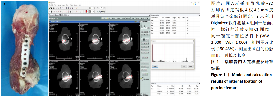
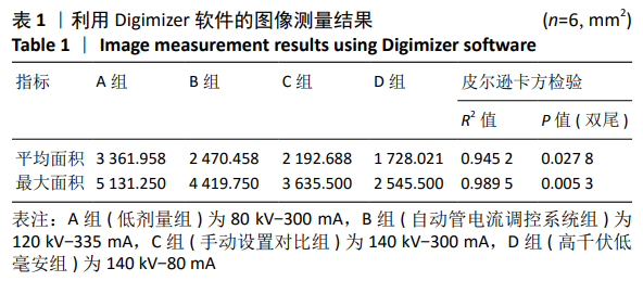
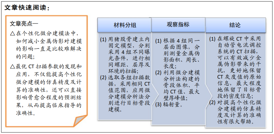
方法:用猪股骨建立内固定模型,分别采用80 kV-300 mA(A组-低剂量组)、120 kV-335 mA(B组-自动管电流调控系统组)、140 kV-300 mA(C组-手动设置对比组)、140 kV-80 mA(D组-高千伏低毫安组)4组不同曝光条件,进行相同螺距、层厚及环境的扫描,采用相同CT值范围,应用微分建模分析法分别进行目标骨段建模。对CT扫描图像中金属伪影的平均面积、最大面积,微分建模分析后的平均CT值、体积和最大壁厚峰值,以及4组扫描条件下辐射量进行比较,判断减小金属伪影、建模准确度及辐射情况,从而选出最优CT扫描参数。
结果与结论:①伪影测量方法结果:A组金属伪影较多,对骨组织遮挡明显,存在漏诊可能;D组金属伪影最少,骨组织周围遮挡少,但图像对比度欠佳,灰雾度较大;而B、C组区别在于图像清晰度,对诊断的准确性基本一致,所以伪影大小排序为:A组>B组>C组>D组;②微分建模分析法结果:B组由于伪影较小,CT值丢失较少,所以仿真出的模型更加接近真实,而且B组采用自动管电流调控系统后,可以明显看出辐射量较小,更加保护患者;③提示B组条件下的CT扫描,可以有效减少金属伪影带来的干扰,更好地保留CT灰度值的原始信息,最大程度地保留目标骨段的密度信息。所以在建立微分建模时,CT自动管电流调控系统为最优的CT扫描参数,不仅可提高个性化微分建模的仿真精度,也增加了计算的准确性。

https://orcid.org/0000-0002-8116-735X (袁新平) 
中国组织工程研究杂志出版内容重点:人工关节;骨植入物;脊柱;骨折;内固定;数字化骨科;组织工程

关键词: 骨愈合, 骨不连, 下肢长骨, 金属伪影, CT值, 微分建模, 内固定

Abstract: BACKGROUND: CT scan and differential modeling are used to analyze the fracture end, which is an effective method to judge the degree of bone healing. To obtain the high precision of differential modeling and simulation, how to select the optimal CT scanning parameters needs further research and analysis. 
OBJECTIVE: To compare the effects of different CT scanning parameters on modeling accuracy in personalized differential modeling analysis, to verify the accuracy and effectiveness of personalized differential modeling in the reduction of simulated target bone segments, and to explore the research value of this method in judging the degree of bone healing of long canals of lower extremities.
METHODS: The model of internal fixation was established with porcine femur. Four groups of 80 kV-300 mA (group A-low dose), 120 kV-335 mA (group B-automatic tube current control system), 140 kV-300 mA (group C-manual setting comparison) and 140 kV-80 mA (group D-high kV and low mA) were used to scan the same object with the same pitch, slice thickness and environment. The scanning data of each group were selected, the same CT value range was used, and the differential modeling analysis method was used to model the target bone segment. The average area and maximum area of metal artifacts in CT scanning images, the average CT value, volume and maximum wall thickness peak after differential modeling analysis, the radiation amount under four groups of scanning conditions were compared to determine the reduction of metal artifacts, modeling accuracy and radiation, so as to select the optimal CT scanning parameters.  
RESULTS AND CONCLUSION: (1) Artifact measurement method results: In group A, there were many metal artifacts, which obviously obstructed bone tissue, and had a possibility of missed diagnosis. In group D, there were minimal metal artifacts, less occlusion around bone tissue, but poor image contrast and high fog. The difference between group B and group C lay in the clarity of images, and the accuracy of diagnosis was basically the same. Therefore, the order of artifact size was as follows: group A > group B > group C > Group D. (2) Differential modeling analysis results: In group B, because of the smaller artifact and less loss of CT value, the simulated model was more close to the reality. Moreover, group B adopted the automatic tube current control system, which could obviously show that the radiation amount was smaller and more protective for patients. (3) It is confirmed that the CT scan under the condition of group B can effectively reduce the interference caused by metal artifacts, better retain the original information of CT gray value, and retain the density information of the target bone segment to the maximum extent. Therefore, when establishing differential modeling, the CT automatic tube current control system is used as the optimal CT scanning parameter, which not only improves the simulation accuracy of personalized differential modeling, but also increases the accuracy of calculation.

Key words: bone healing, nonunion, lower limb long bone, metal artifact, CT value, differential modeling, internal fixation

中图分类号: