中国组织工程研究 ›› 2019, Vol. 23 ›› Issue (18): 2888-2893.doi: 10.3969/j.issn.2095-4344.1713

• 纳米生物材料 nanobiomaterials • 上一篇    下一篇

骨碎补总黄酮联合纳米骨材料诱导MC3T3-E1细胞的成骨分化及其机制

李晋玉1,俞  兴1,姜俊杰2,徐  林1,赵学千1,孙  旗1,郑晨颖1,白春晓1,刘楚吟1,张  喆1,贾育松1
  

  1. 1北京中医药大学东直门医院骨科,北京市  100700;2中国中医科学院中医临床基础医学研究所,北京市  100700
  • 收稿日期:2019-01-22 出版日期:2019-06-28 发布日期:2019-06-28
  • 通讯作者: 贾育松,主任医师,北京中医药大学东直门医院骨一科,北京市 100700
  • 作者简介:李晋玉,男,1984年生,山西省晋城市人,汉族,2018年北京中医药大学毕业,博士,副主任医师,主要从事中医药在骨组织工程中的应用研究。
  • 基金资助:

    国家自然科学基金项目(81503601),项目负责人:李晋玉;国家自然科学基金项目(81603517),项目负责人:姜俊杰;北京中医药大学东直门医院青苗人才项目(DZMYS-201802),项目负责人:李晋玉

Osteogenic differentiation of MC3T3-E1 cells induced by osteopractic total flavone and nano-hydroxyapatite/collagen composite and the underlying mechanism

Li Jinyu1, Yu Xing1, Jiang Junjie2, Xu Lin1, Zhao Xueqian1, Sun Qi1, Zheng Chenying1, Bai Chunxiao1, Liu Chuyin1, Zhang Zhe1, Jia Yusong1 
  

  1. 1Department of Orthopedics, Dongzhimen Hospital, Beijing University of Chinese Medicine, Beijing 100700, China; 2Institute for Basic Research in Clinical Medicine, China Academy of Chinese Medical Sciences, Beijing 100700, China
  • Received:2019-01-22 Online:2019-06-28 Published:2019-06-28
  • Contact: Jia Yusong, Chief physician, Department of Orthopedics, Dongzhimen Hospital, Beijing University of Chinese Medicine, Beijing 100700, China
  • About author:Li Jinyu, MD, Associate chief physician, Department of Orthopedics, Dongzhimen Hospital, Beijing University of Chinese Medicine, Beijing 100700, China
  • Supported by:

    the National Natural Science Foundation of China, No. 81503601 (to LJY) and 81603517 (to JJJ); the Young Talent Project of Dongzhimen Hospital of Beijing University of Chinese Medicine, No. DZMYS-201802 (to LJY)

摘要:

文章快速阅读:


文题释义:
骨碎补总黄酮:是由水龙骨科植物槲蕨干燥根茎中提取的有效成分,实验证明骨碎补总黄酮能够促进成骨细胞的增殖,抑制破骨细胞的成熟分化。
骨桥蛋白:一种含有Arg-Gly-Asp(RGD)结构的酸性糖蛋白,存在于矿化和活性沉积区,它与诱导成骨细胞成熟表型表达和矿化骨基质形成的活性蛋白密切相关;在成矿阶段,骨连接素、纤连蛋白与骨桥蛋白的表达高度相关。
 
 
背景:前期研究发现,骨碎补总黄酮可促进纳米骨材料表面MC3T3-E1细胞的成骨分化,其作用机制有待进一步研究。
目的:探讨骨碎补总黄酮联合纳米骨材料对MC3T3-E1细胞成骨分化的影响及作用机制。
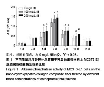
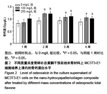
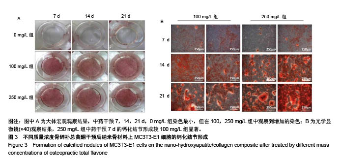
方法:将MC3T3-E1细胞接种于纳米骨材料表面,分别加入含0,100,250 mg/L骨碎补总黄酮的培养基培养。培养第1,3,5,7,9,11,14天,检测细胞碱性磷酸酶活性;培养第1,2,3,4周,ELISA法检测培养上清中骨钙蛋白水平;培养第7,14,21天,茜素红S染色观察细胞钙化结节形成情况;培养第14天,PCR检测成骨相关基因表达。

结果与结论:①第1,3,14天,3组细胞碱性磷酸酶活性比较差异无显著性意义(P > 0.05);培养第5,7,9天,100,250 mg/L组碱性磷酸酶活性均高于0 mg/L组(P < 0.05),100 mg/L组、250 mg/L组间比较差异无显著性意义(P > 0.05);②培养第1,2,3,4周,100,250 mg/L组骨钙蛋白质量浓度均高于0 mg/L组       (P < 0.05),100 mg/L组、250 mg/L组间比较差异无显著性意义(P > 0.05);③培养第7,14,21天,100,250 mg/L组可见明显的钙化结节;④培养第14天,100,250 mg/L组骨钙蛋白、Ⅰ型胶原和骨桥蛋白mRNA表达水平均高于0 mg/L组(P < 0.05),100 mg/L组、250 mg/L组间骨钙蛋白、Ⅰ型胶原和骨桥蛋白mRNA表达比较差异无显著性意义(P > 0.05);⑤结果表明,骨碎补总黄酮联合纳米骨材料可诱导MC3T3-E1细胞分化为成骨细胞,其机制可能是通过提高碱性磷酸酶活性、骨钙蛋白水平及Ⅰ型胶原、骨钙蛋白和骨桥蛋白mRNA表达来实现的。

关键词: 纳米骨材料, 骨碎补总黄酮, MC3T3-E1细胞, 成骨分化, 骨桥蛋白, 骨钙蛋白;, 国家自然科学基金

Abstract:

BACKGROUND: Preliminary study has found that osteopractic total flavone can promote osteogenic differentiation of MC3T3-E1 cells on the surface of nano-hydroxyapatite/collagen composite, but the underlying mechanism needs to be studied in depth.
OBJECTIVE: To investigate the effect of osteopractic total flavone combined with nano-hydroxyapatite/collagen composite on the osteogenic differentiation of MC3T3-E1 cells and its mechanism.
METHODS: MC3T3-E1 cells were seeded on the surface of nano-hydroxyapatite/collagen composite and cultured in culture media containing 0, 100 and 250 mg/L osteopractic total flavone, respectively. After 1, 3, 5, 7, 9, 11 and 14 days of culture, the activity of alkaline phosphatase was detected. After 1, 2, 3 and 4 weeks of culture, the level of osteocalcin in the culture supernatant was detected by ELISA. Alizarin red staining was used to observe the formation of calcified nodules at 7, 14 and 21 days of culture. At 14 days of culture, the expression of osteogenic related genes was detected by PCR.
RESULTS AND CONCLUSION: (1) There was no significant difference in the alkaline phosphatase activity at 1, 3 and 14 days among groups (P > 0.05). At 5, 7 and 9 days of culture, the alkaline phosphatase activity in the 100 and 250 mg/L groups was higher than that in the 0 mg/L group (P < 0.05), and there was no significant difference between 100 and 250 mg/L groups (P > 0.05). (2) The mass concentration of osteocalcin in the 100 and 250 mg/L groups was higher than that in the 0 mg/L group at 1, 2, 3 and 4 weeks of culture (P < 0.05), and there was no significant difference between 100 and 250 mg/L groups (P > 0.05). (3) At 7, 14 and 21 days of culture, obvious calcified nodules were visible in the 100 and 250 mg/L groups. (4) At 14 days of culture, the mRNA expression levels of osteocalcin, type I collagen and osteopontin in the 100 and 250 mg/L groups were higher than that in the 0 mg/L group (P < 0.05), and there was no significant difference between 100 and 250 mg/L groups (P > 0.05). (5) These results imply that osteopractic total flavone combined with nano-hydroxyapatite/collagen composite can induce MC3T3-E1 cells to differentiate into osteoblasts, which may be by increasing alkaline phosphatase activity, osteocalcin level and mRNA expression of type I collagen, osteocalcin and osteopontin.

Key words: nano-hydroxyapatite/collagen composite, osteopractic total flauone, MC3T3-E1 cells, osteogenic differentiation, osteopontin, osteocalcin, the National Natural Science Foundation of China

中图分类号: