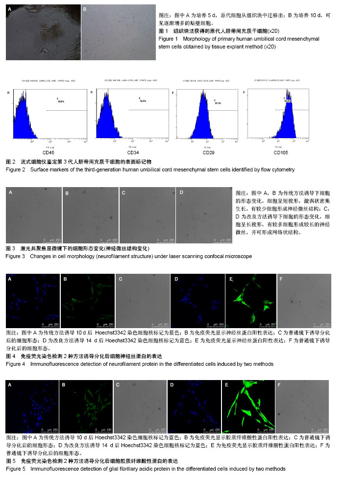
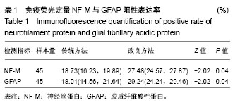
| [1] Pop DM, Sori??u O, ?u?man S, et al. Potential of placental-derived human mesenchymal stem cells for osteogenesis and neurogenesis. Rom J Morphol Embryol. 2015;56(3):989-996. [2] Nitkin CR, Bonfield TL. Concise Review: Mesenchymal Stem Cell Therapy for Pediatric Disease: Perspectives on Success and Potential Improvements. Stem Cells Transl Med. 2017;6(2):539-565. [3] Nekanti U, Mohanty L, Venugopal P, et al. Optimization and scale-up of Wharton's jelly-derived mesenchymal stem cells for clinical applications. Stem Cell Res. 2010;5(3):244-254. [4] Gao F, Chiu SM, Motan DA, et al. Mesenchymal stem cells and immunomodulation: current status and future prospects. Cell Death Dis. 2016;7:e2062. [5] Du WJ, Chi Y, Yang ZX, et al. Heterogeneity of proangiogenic features in mesenchymal stem cells derived from bone marrow, adipose tissue, umbilical cord, and placenta. Stem Cell Res Ther. 2016;7(1):163. [6] Antunes MA, Laffey JG, Pelosi P, et al. Mesenchymal stem cell trials for pulmonary diseases. J Cell Biochem. 2014;115(6):1023-1032. [7] Latifpour M, Shakiba Y, Amidi F, et al. Differentiation of human umbilical cord matrix-derived mesenchymal stem cells into germ-like cells. Avicenna J Med Biotechnol. 2014;6(4):218-227. [8] Tao R, Sun TJ, Han YQ, et al. Epimorphin-induced differentiation of human umbilical cord mesenchymal stem cells into sweat gland cells. Eur Rev Med Pharmacol Sci. 2014;18(9):1404-1410. [9] Li W, Ye B, Cai XY, et al. Differentiation of human umbilical cord mesenchymal stem cells into prostate-like epithelial cells in vivo. PLoS One. 2014;9(7):e102657. [10] Bai J, Hu Y, Wang YR, et al. Comparison of human amniotic fluid-derived and umbilical cord Wharton's Jelly-derived mesenchymal stromal cells: Characterization and myocardial differentiation capacity. J Geriatr Cardiol. 2012;9(2):166-171. [11] Hu Y, Liang J, Cui H, et al. Wharton's jelly mesenchymal stem cells differentiate into retinal progenitor cells. Neural Regen Res. 2013; 8(19):1783-1792. [12] Hang H, Yu Y, Wu N, et al. Induction of highly functional hepatocytes from human umbilical cord mesenchymal stem cells by HNF4α transduction. PLoS One. 2014;9(8):e104133. [13] Qu H, Liu X, Ni Y, et al. Laminin 411 acts as a potent inducer of umbilical cord mesenchymal stem cell differentiation into insulin-producing cells. J Transl Med. 2014;12:135. [14] Doan CC, Le TL, Hoang NS, et al. Differentiation of umbilical cord lining membrane-derived mesenchymal stem cells into endothelial-like cells. Iran Biomed J. 2014;18(2):67-75. [15] Paldino E, Cenciarelli C, Giampaolo A, et al. Induction of dopaminergic neurons from human Wharton's jelly mesenchymal stem cell by forskolin. J Cell Physiol. 2014;229(2):232-244. [16] Alves da Silva ML, Costa-Pinto AR, Martins A, et al. Conditioned medium as a strategy for human stem cells chondrogenic differentiation. J Tissue Eng Regen Med. 2015;9(6):714-723. [17] Ali H, Al-Yatama MK, Abu-Farha M, et al. Multi-lineage differentiation of human umbilical cord Wharton's Jelly Mesenchymal Stromal Cells mediates changes in the expression profile of stemness markers. PLoS One. 2015;10(4):e0122465. [18] Bárcia RN, Santos JM, Filipe M, et al. What Makes Umbilical Cord Tissue-Derived Mesenchymal Stromal Cells Superior Immunomodulators When Compared to Bone Marrow Derived Mesenchymal Stromal Cells. Stem Cells Int. 2015;2015:583984. [19] Hu SL, Luo HS, Li JT, et al. Functional recovery in acute traumatic spinal cord injury after transplantation of human umbilical cord mesenchymal stem cells. Crit Care Med. 2010;38(11):2181-2189. [20] Pang KM, Sung MA, Alrash-dan MS, et al. Tans-plantation of mesenchymal stem cells from human umbilical cord versus human umbilical cord blood for peripheral nerve regeneration. Neural Regen Res. 2010;5(11):838-845. [21] Cheng H, Liu X, Hua R, et al. Clinical observation of umbilical cord mesenchymal stem cell transplantation in treatment for sequelae of thoracolumbar spinal cord injury. J Transl Med. 2014;12:253. [22] Chen J, Venkat P, Zacharek A, et al. Neurorestorative therapy for stroke. Front Hum Neurosci. 2014;8:382. [23] 张飞,王一雄,武忠炎,等.人脐带间充质干细胞生物特性比较:胰酶冷消化和组织块法体外培养[J].中国组织工程研究, 2014,18(41):6614-6619.[24] McElreavey KD, Irvine AI, Ennis KT, et al. Isolation, culture and characterisation of fibroblast-like cells derived from the Wharton's jelly portion of human umbilical cord. Biochem Soc Trans. 1991;19(1):29S. [25] 王武,张飞,李贵才,等.以 EdU 体外标记人脐带间充质干细胞:5,10 μmol/L 是其最适浓度[J].中国组织工程研究, 2015,19(32):5167-5171.[26] Ianus A, Holz GG, Theise ND, et al. In vivo derivation of glucose-competent pancreatic endocrine cells from bone marrow without evidence of cell fusion. J Clin Invest. 2003;111(6):843-850. [27] Ra JC, Shin IS, Kim SH, et al. Safety of intravenous infusion of human adipose tissue-derived mesenchymal stem cells in animals and humans. Stem Cells Dev. 2011;20(8):1297-1308. [28] Nazarov I, Lee JW, Soupene E, et al. Multipotent stromal stem cells from human placenta demonstrate high therapeutic potential. Stem Cells Transl Med. 2012;1(5):359-372. [29] Rohban R, Pieber TR. Mesenchymal Stem and Progenitor Cells in Regeneration: Tissue Specificity and Regenerative Potential. Stem Cells Int. 2017;2017:5173732. [30] Guan LX, Guan H, Li HB, et al. Therapeutic efficacy of umbilical cord-derived mesenchymal stem cells in patients with type 2 diabetes. Exp Ther Med. 2015;9(5):1623-1630. [31] Zhao XF, Xu Y, Zhu ZY, et al. Clinical observation of umbilical cord mesenchymal stem cell treatment of severe systolic heart failure. Genet Mol Res. 2015;14(2):3010-3017. [32] D'souza N, Rossignoli F, Golinelli G, et al. Mesenchymal stem/stromal cells as a delivery platform in cell and gene therapies. BMC Med. 2015;13:186. [33] Hendijani F, Sadeghi-Aliabadi H, Haghjooy Javanmard S. Comparison of human mesenchymal stem cells isolated by explant culture method from entire umbilical cord and Wharton's jelly matrix. Cell Tissue Bank. 2014;15(4):555-565. [34] Zhou X, Gu J, Gu Y, et al. Human Umbilical Cord-Derived Mesenchymal Stem Cells Improve Learning and Memory Function in Hypoxic-Ischemic Brain-Damaged Rats via an IL-8-Mediated Secretion Mechanism Rather than Differentiation Pattern Induction. Cell Physiol Biochem. 2015;35(6):2383-2401. [35] Álvarez D, Levine M, Rojas M. Regenerative medicine in the treatment of idiopathic pulmonary fibrosis: current position. Stem Cells Cloning. 2015;8:61-65. [36] Rismanchi N, Floyd CL, Berman RF, et al. Cell death and long-term maintenance of neuron-like state after differentiation of rat bone marrow stromal cells: a comparison of protocols. Brain Res. 2003;991(1-2):46-55. [37] 孙丽,于丽,张华芳,等.人脐带间充质干细胞的富集及向神经细胞的诱导分化[J].神经解剖杂志,2011,27(2):185-190.[38] 郜元军,钱伟,汪涉海,等.大鼠骨髓基质干细胞诱导分化为肠神经细胞的体外研究[J].中华消化杂志,2006,26(10):661-665. |
.jpg)


.jpg)