| [1] Li P, Bai Y, Yin G, et al. Synergistic and sequential effects of BMP-2, bFGF and VEGF on osteogenic differentiation of rat osteoblasts.J Bone Miner Metab. 2014;32(6):627-635.
[2] Barati D, Shariati SRP, Moeinzadeh S, et al. Spatiotemporal release of BMP-2 and VEGF enhances osteogenic and vasculogenic differentiation of human mesenchymal stem cells and endothelial colony-forming cells co-encapsulated in a patterned hydrogel.J Control Release. 2016;223:126-136.
[3] Nakano K, Murata K, Omokawa S, et al. Promotion of Osteogenesis and Angiogenesis in Vascularized Tissue-Engineered Bone Using Osteogenic Matrix Cell Sheets.Plast Reconstr Surg. 2016;137(5):1476-1484.
[4] Shen Y, Qiao H, Fan Q, et al. Potentiated Osteoinductivity via Cotransfection with BMP-2 and VEGF Genes in Microencapsulated C2C12 Cells.Biomed Res Int. 2015;2015: 435253.
[5] Kim IS, Song YM, Hwang SJ.Osteogenic responses of human mesenchymal stromal cells to static stretch.J Dent Res. 2010; 89(10):1129-1134.
[6] Taghiyar L, Hosseini S, Hesaraki M, et al. Isolation, Characterization and Osteogenic Potential of Mouse Digit Tip Blastema Cells in Comparison with Bone Marrow-Derived Mesenchymal Stem Cells In Vitro.Cell J. 2018;19(4):585-598.
[7] Bakopoulou A, Apatzidou D, Aggelidou E, et al. Isolation and prolonged expansion of oral mesenchymal stem cells under clinical-grade, GMP-compliant conditions differentially affects "stemness" properties.Stem Cell Res Ther. 2017;8(1):247.
[8] Shokohi R, Nabiuni M, Irian S, et al. In Vitro Effects of Wistar Rat Prenatal and Postnatal Cerebrospinal Fluid on Neural Differentiation and P roliferation of Mesenchymal Stromal Cells Derived from Bone Marrow.Cell J. 2018;19(4):537-544.
[9] Shi XL, Hu BB, Ren MM, et al. Hypoxia regulates the expression of OPG/RANKL mRNA in rat bone marrow mesenchymal stem cells.Shanghai Kou Qiang Yi Xue. 2017; 26(3):258-262.
[10] Lin J, Shao J, Juan L, et al. Enhancing bone regeneration by combining mesenchymal stem cell sheets with β-TCP/COL-I scaffolds.J Biomed Mater Res B Appl Biomater. 2018;106(5): 2037-2045.
[11] He XT, Li X, Yin Y, et al. The effects of conditioned media generated by polarized macrophages on the cellular behaviours of bone marrow mesenchymal stem cells.J Cell Mol Med. 2018;22(2):1302-1315.
[12] Tirkkonen L, Haimi S, Huttunen S, et al. Osteogenic medium is superior to growth factors in differentiation of human adipose stem cells towards bone-forming cells in 3D culture. Eur Cell Mater. 2013;25:144-158.
[13] Jiang J, Fan CY, Zeng BF.Osteogenic differentiation effects on rat bone marrow-derived mesenchymal stromal cells by lentivirus-mediated co-transfection of human BMP2 gene and VEGF165 gene.Biotechnol Lett. 2008;30(2):197-203.
[14] Li H, Li J, Jiang J, et al. An osteogenesis/ angiogenesis-stimulation artificial ligament for anterior cruciate ligament reconstruction.Acta Biomater. 2017;54: 399-410.
[15] Perez RA, Kim JH, Buitrago JO, et al. Novel therapeutic core-shell hydrogel scaffolds with sequential delivery of cobalt and bone morphogenetic protein-2 for synergistic bone regeneration.Acta Biomater. 2015;23:295-308.
[16] Herath TDK, Larbi A, Teoh SH, et al. Neutrophil-mediated enhancement of angiogenesis and osteogenesis in a novel triple cell co-culture model with endothelial cells and osteoblasts.J Tissue Eng Regen Med. 2018;12(2): e1221-e1236.
[17] Liu S, Jin D, Wu JQ, et al. Neuropeptide Y stimulates osteoblastic differentiation and VEGF expression of bone marrow mesenchymal stem cells related to canonical Wnt signaling activating in vitro.Neuropeptides. 2016;56:105-113.
[18] Kumar S, Wan C, Ramaswamy G, et al. Mesenchymal stem cells expressing osteogenic and angiogenic factors synergistically enhance bone formation in a mouse model of segmental bone defect.Mol Ther. 2010;18(5):1026-1034.
[19] Fu TS, Chang YH, Wong CB, et al. Mesenchymal stem cells expressing baculovirus-engineered BMP-2 and VEGF enhance posterolateral spine fusion in a rabbit model.Spine J. 2015;15(9):2036-2044.
[20] Wang W, Kratz K, Behl M, et al. The interaction of adipose-derived human mesenchymal stem cells and polyether ether ketone.Clin Hemorheol Microcirc. 2015;61(2): 301-321.
[21] Han TY, Liu XW, Liang N, et al. In vitro effects of recombinant adenovirus-mediated bone morphogenetic protein 2/vascular endothelial growth factor 165 on osteogenic differentiation of bone marrow mesenchymal stem cells.Artif Cells Nanomed Biotechnol. 2017;45(1):108-114.
[22] Lin Z, Wang JS, Lin L, et al. Effects of BMP2 and VEGF165 on the osteogenic differentiation of rat bone marrow-derived mesenchymal stem cells.Exp Ther Med. 2014;7(3):625-629.
[23] Kanczler JM, Ginty PJ, White L, et al. The effect of the delivery of vascular endothelial growth factor and bone morphogenic protein-2 to osteoprogenitor cell populations on bone formation.Biomaterials. 2010;31(6):1242-1250.
[24] Tian XB, Sun L, Yang SH, et al. Ectopic osteogenesis of mouse bone marrow stromal cells transfected with BMP 2/VEGF(165) genes in vivo.Orthop Surg. 2009;1(4):322-325.
[25] Kärner E, Bäckesjö CM, Cedervall J, et al. Dynamics of gene expression during bone matrix formation in osteogenic cultures derived from human embryonic stem cells in vitro.Biochim Biophys Acta. 2009;1790(2):110-118.
[26] Janko M, Sahm J, Schaible A, et al. Comparison of three different types of scaffolds preseeded with human bone marrow mononuclear cells on the bone healing in a femoral critical size defect model of the athymic rat.J Tissue Eng Regen Med. 2018;12(3):653-666.
[27] Farhadi J, Jaquiery C, Barbero A, et al. Differentiation-dependent up-regulation of BMP-2, TGF-beta1, and VEGF expression by FGF-2 in human bone marrow stromal cells.Plast Reconstr Surg. 2005;116(5):1379-1386.
[28] Curtin CM, Tierney EG, McSorley K, et al. Combinatorial gene therapy accelerates bone regeneration: non-viral dual delivery of VEGF and BMP2 in a collagen-nanohydroxyapatite scaffold.Adv Healthc Mater. 2015;4(2):223-227.
[29] Xiao C, Zhou H, Liu G, et al. Bone marrow stromal cells with a combined expression of BMP-2 and VEGF-165 enhanced bone regeneration.Biomed Mater. 2011;6(1):015013.
[30] Song X, Liu S, Qu X, et al. BMP2 and VEGF promote angiogenesis but retard terminal differentiation of osteoblasts in bone regeneration by up-regulating Id1.Acta Biochim Biophys Sin (Shanghai). 2011;43(10):796-804.
[31] Lin CY, Chang YH, Kao CY, et al. Augmented healing of critical-size calvarial defects by baculovirus-engineered MSCs that persistently express growth factors.Biomaterials. 2012;33(14):3682-3692.
[32] Zhang C, Yu L, Liu S, et al. Human amnion-derived mesenchymal stem cells promote osteogenic and angiogenic differentiation of human adipose-derived stem cells.PLoS One. 2017;12(10):e0186253.
[33] 胡正雄,李彪,蓝天.TGF-β2和geneX对BrdU标记骨髓间充质干细胞增殖与成骨分化的作用[J].昆明医科大学学报, 2016,37(2): 10-14.
[34] 张爱军,闫志勇.TGF-β对创伤愈合与瘢痕形成的影响及中药的干预作用[J].西北药学杂志,2013,16(1):101-105. |
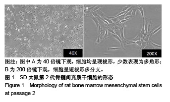
.jpg)
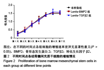
.jpg)
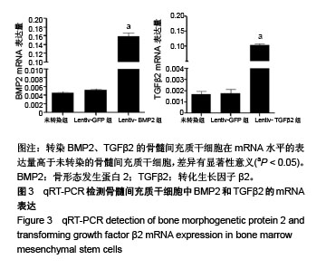
.jpg)