中国组织工程研究 ›› 2019, Vol. 23 ›› Issue (35): 5638-5644.doi: 10.3969/j.issn.2095-4344.1429
• 组织构建基础实验 basic experiments in tissue construction • 上一篇 下一篇
shRNA沉默骨膜蛋白基因抑制人骨肉瘤的血管生成
徐朝健,张 龙,程才统,冯 毅,吕 嘉,孙晓娟,吕 智
- (山西医科大学第二医院骨科,山西省太原市 030001)
Silencing of periosteal protein gene by shRNA inhibits angiogenesis in human osteosarcoma
Xu Chaojian, Zhang Long, Cheng Caitong, Feng Yi, Lü Jia, Sun Xiaojuan, Lü Zhi
- (Department of Orthopedics, the Second Hospital of Shanxi Medical University, Taiyuan 030001, Shanxi Province, China)
摘要:
文章快速阅读:
.jpg) 文题释义:
骨膜蛋白:又称成骨细胞特异性因子2,是一种细胞外基质蛋白,参与细胞的募集、黏附过程,并且在多种恶性肿瘤中呈现高表达,与肿瘤的迁移、增殖和血管生成能力有关。
小动物活体荧光断层成像技术:是小动物活体成像领域的新一代技术,利用萤火虫荧光素酶或荧光蛋白作为报告基因,通过转基因技术体外标记肿瘤细胞而直接观测肿瘤的发展变化,或者标记特定基因研究肿瘤相关基因在肿瘤发生发展中的作用。
文题释义:
骨膜蛋白:又称成骨细胞特异性因子2,是一种细胞外基质蛋白,参与细胞的募集、黏附过程,并且在多种恶性肿瘤中呈现高表达,与肿瘤的迁移、增殖和血管生成能力有关。
小动物活体荧光断层成像技术:是小动物活体成像领域的新一代技术,利用萤火虫荧光素酶或荧光蛋白作为报告基因,通过转基因技术体外标记肿瘤细胞而直接观测肿瘤的发展变化,或者标记特定基因研究肿瘤相关基因在肿瘤发生发展中的作用。
文题释义:
骨膜蛋白:又称成骨细胞特异性因子2,是一种细胞外基质蛋白,参与细胞的募集、黏附过程,并且在多种恶性肿瘤中呈现高表达,与肿瘤的迁移、增殖和血管生成能力有关。
小动物活体荧光断层成像技术:是小动物活体成像领域的新一代技术,利用萤火虫荧光素酶或荧光蛋白作为报告基因,通过转基因技术体外标记肿瘤细胞而直接观测肿瘤的发展变化,或者标记特定基因研究肿瘤相关基因在肿瘤发生发展中的作用。摘要
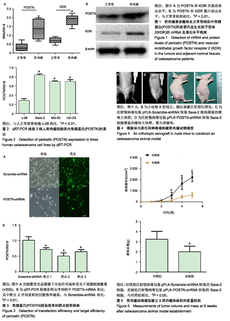
背景:骨膜蛋白(periostin,POSTN)是一种细胞外基质蛋白,在多种肿瘤组织中表达上调,与肿瘤的迁移、增殖和血管生成能力有关。血管内皮生长因子受体2(vascular endothelial growth factor2,VEGFR2/KDR)与其配体血管内皮生长因子结合后主要调控血管的发生和构建,但KDR与POSTN在骨肉瘤中的相互作用尚不清楚。
目的:探讨POSTN在体内、外水平对人骨肉瘤增殖和体内血管生成能力影响及其作用机制。
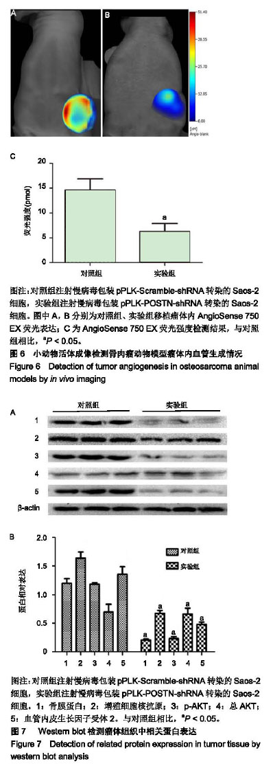
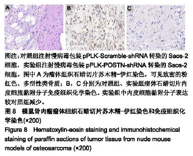
方法:①体外实验:qRT-PCR和Western blot检测人骨肉瘤临床标本组织中POSTN和KDR的mRNA及蛋白表达水平;qRT-PCR检测3株不同人骨肉瘤细胞系内POSTN表达,筛选基因表达最高的细胞系进行转染实验;转染慢病毒包装pPLK-POSTN-shRNA进入Saos-2细胞,选出3个作用靶点中抑制率最高的序列进行后续实验;②体内实验:取裸鼠(购自北京维通利华实验动物技术有限公司)12只,实验组(n=6)将慢病毒包装pPLK-POSTN-shRNA转染的Saos-2细胞注射至胫骨髓腔中,对照组(n=6)将慢病毒包装pPLK-Scramble-shRNA转染的Saos-2细胞注射至胫骨髓腔中,接种5周后,测量瘤体体积与质量;接种3周后,应用小动物活体成像检测血管生成情况;接种5周后取瘤体组织,Western blot检测POSTN、增殖细胞核抗原、KDR和p-AKT蛋白表达,病理切片免疫组织化学染色内皮细胞黏附分子蛋白表达。实验方案经山西医科大学第二医院伦理委员会批准(编号:2018002)。
结果与结论:①体外实验:骨肉瘤组织中POSTN和KDR的mRNA及蛋白水平表达均高于正常骨组织(P < 0.01);Saos-2细胞中的POSTN基因表达量较MG-63和U2-OS细胞高;②体内实验:实验组瘤体体积和质量均小于对照组(P < 0.05);实验组瘤体内血管生成量少于对照组(P < 0.05);实验组POSTN、增殖细胞核抗原、KDR和p-AKT及内皮细胞黏附分子蛋白表达均低于对照组(P < 0.05);③结果表明:沉默POSTN基因可抑制骨肉瘤血管生成,其机制可能与KDR/PI3K/AKT通路活化有关。
中图分类号:
.jpg) 文题释义:
骨膜蛋白:又称成骨细胞特异性因子2,是一种细胞外基质蛋白,参与细胞的募集、黏附过程,并且在多种恶性肿瘤中呈现高表达,与肿瘤的迁移、增殖和血管生成能力有关。
小动物活体荧光断层成像技术:是小动物活体成像领域的新一代技术,利用萤火虫荧光素酶或荧光蛋白作为报告基因,通过转基因技术体外标记肿瘤细胞而直接观测肿瘤的发展变化,或者标记特定基因研究肿瘤相关基因在肿瘤发生发展中的作用。
文题释义:
骨膜蛋白:又称成骨细胞特异性因子2,是一种细胞外基质蛋白,参与细胞的募集、黏附过程,并且在多种恶性肿瘤中呈现高表达,与肿瘤的迁移、增殖和血管生成能力有关。
小动物活体荧光断层成像技术:是小动物活体成像领域的新一代技术,利用萤火虫荧光素酶或荧光蛋白作为报告基因,通过转基因技术体外标记肿瘤细胞而直接观测肿瘤的发展变化,或者标记特定基因研究肿瘤相关基因在肿瘤发生发展中的作用。