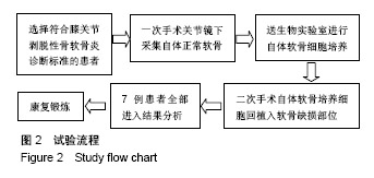
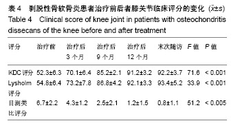
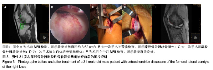
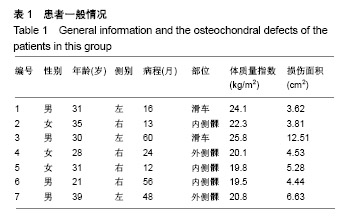
| [1]Chadli L, Cottalorda J, Delpont M,et al. Autologous osteochondral mosaicplasty in osteochondritis dissecans of the patella in adolescents.Int Orthop.2017;41(1):197-202.[2]吕帅洁,毛强,童培建,等.剥脱性骨软骨炎的研究进展[J].中国骨伤,2014,27(9):787-791.[3]Weiss JM,Shea KG,Jacobs JC,et al.Incidence of Osteochondritis Dissecans in Adults.Am J Sports Med. 2018;46(7):1592-1595.[4]黄迅悟,冯会成,孙继桐,等.成年人股骨髁剥脱性骨软骨炎的外科治疗[J].中华关节外科杂志(电子版), 2013,7(6):747-750.[5]Kirsch JM,Thomas JR,Khan M,et al.Return to Play After Osteochondral Autograft Transplantation of the Capitellum: A SystematicReview.Arthroscopy.2017;33(7):1412-1420.e1. [6]Peterson L,Menche D,Grande D,et al.Chondrocyte transplatation. An experimental model in the rabbit.Trans Orthop Res Soc.1984;9:218.[7]Brittberg M,Lindahl A,Nilsson A,et al.Treatment of deep cartilage defects in the knee with autologous chondrocyte transplantation.N Engl J Med.1994;331(14):889-895.[8]Schneider U,Rackwitz L,Andereya S,et al.A prospective multicenter study on the outcome of type I collagen hydrogel-based autologous chondrocyte implantation (CaReS) for the repair of articular cartilage defects in the knee.Am J Sports Med. 2011;39(2):2558-2565.[9]Rackwitz L,Schneider U,Andereya S,et al.Reconstruction of osteochondral defects with a collagen I hydrogel. Results of a prospective multicenter study. Orthopade. 2012;41(4): 268-279.[10]Kocher MS,Steadman JR,Briggs KK,et al.Reliability, Validity, and Responsiveness of the Lysholm Knee Scale for Various Chondral Disorders of the Knee.J Bone Joint Surg Am. 2004;86-A(6):1139-1145.[11]Collins NJ,Misra D,Felson DT,et al.Measures of knee function: International Knee Documentation Committee (IKDC) Subjective Knee Evaluation Form, Knee Injury and Osteoarthritis Outcome Score (KOOS), Knee Injury and Osteoarthritis Outcome Score Physical Function Short Form (KOOS-PS), Knee Outcome Survey Activities of Daily Living Scale (KOS-ADL), Lysholm Knee Scoring Scale, Oxford Knee Score (OKS), Western Ontario and McMaster Universities Osteoarthritis Index (WOMAC), Activity Rating Scale (ARS), and Tegner Activity Score (TAS).Arthritis Care Res (Hoboken). 2011;63 Suppl 11:S208-228.[12]刘志军,胡兵伟.预防老年患者麻醉并发症管理措施的效果观察[J].医院管理论坛,2018,32(5):15-16.[13]Parikh SN,Allen M,Wall EJ,et al.The reliability to determine healing in osteochondritis dissecans from radiographic assessment.J Pediatr Orthop.2012;32:e35-39.[14]陈清华,陈士明,于建农.剥脱性骨软骨炎治疗研究进展[J].浙江临床医学,2017,19(1):183-185.[15]Meyer LF.Research in Osteochondritis Dissecans of the Knee: 2016 Update.J Knee Surg. 2016;29(7):533-538.[16]Chadli L,Cottalorda J,Delpont M,et al.Autologous osteochondral mosaicplasty in osteochondritis dissecans of the patella in adolescents.Int Orthop.2017;41(1):197-202.[17]Logli AL,Bernard CD,O'Driscoll SW,et al. Osteochondritis dissecans lesions of the capitellum in overhead athletes: a review of current evidence and proposed treatment algorithm.Curr Rev Musculoskelet Med. 2019;12(1):1-12.[18]Bray CC, Watson ST. Current review of juvenile osteochondritis dissecans of the knee. Curr Orthop Pract.2015;26(5):466-474[19]Finnøy A, Olstad K, Lilledahl MB.Characterization of cellular and matrix alterations in the early pathogenesis of osteochondritis dissecans in pigs using second harmonic generation and two-photon excitation fluorescence microscopy.J Orthop Res. 2018. doi: 10.1002/jor.23874. [Epub ahead of print][20]Pareek A,Carey JL,Reardon PJ,et al.Long-Term Outcomes after Autologous Chondrocyte Implantation: A Systematic Review at Mean Follow-Up of 11.4 Years.Cartilage. 2016; 7(4):298-308.[21]Kraeutler MJ,Belk JW,Purcell JM ,et al.Microfracture Versus Autologous Chondrocyte Implantation for Articular Cartilage Lesions in the Knee: A Systematic Review of 5-Year Outcomes.Am J Sports Med.2018;46(4):995-999.[22]李刚,焦强,王春理,等.基质诱导自体软骨细胞移植治疗软骨损伤的研究现状[J].中国矫形外科杂志,2017,25(14): 1292-1296.[23]安勇,王丹亭,岳宏,等.基质诱导的自体软骨细胞移植治疗膝关节软骨损伤的研究进展[J].医学研究杂志,2017,46(7): 177-180.[24]Schagemann J,Mrosek E,Landers R,et al.Morphology and function of bovine articular cartilage chondrocytes in 3-D hydrogel culture.Cells Tissues Organs.2006;182(2):89-97.[25]Welsch G,Mamisch T,Zak L,et al.Evaluation of cartilage repair tissue after matrix-associated autologous chondrocyte transplantation using a hyaluronic-based or a collagen-based scaffold with morphological MOCART scoring and biochemical T2 mapping: preliminary results. Am J Sports Med.2010;38(5):934-942.[26]张萌,张慎启,邵林.关节软骨缺损的手术治疗[J].现代医学, 2012,40(6):736-739.[27]Krishnan S,Skinner J,Bartlett W,et al.Who is the ideal candidate for autologous chondrocyte implantation.J Bone Joint Surg Br.2006;88(1):61-64.[28]Nawaz S,Bentley G,Briggs T,et al.Autologous Chondrocyte Implantation in the Knee. Mid-Term to Long-Term Results.J Bone Joint Surg Am.2014;96(10):824-830.[29]Biant LC,Mcnicholas MJ,Sprowson AP,et al.The surgical management of symptomatic articular cartilage defects of the knee: Consensus statements from United Kingdom knee surgeons.Knee.2015; 22(5):446-449.[30]Thomas R, Peter M, Elem S, et al. Early resumption of physical activities leads to inferior clinical outcomes after matrix-based autologous chondrocyte implantation in the knee. Knee Surg Sports Traumatol Arthrosc. 2014;22(6): 1345-1352. |
.jpg)

.jpg)