| [1] 夏玲,王欢,窦晓莉,等.膝骨性关节炎的非手术治疗现状[J].中国老年学杂志,2011,4(31):1491-1494.[2] Hootman JM, Helmick CG. Projections of US prevalence of arthritis and associated activity limitations. Arthritis Rheum. 2006;54:226-229. [3] Kurtz SM, Lau E, Ong K, et al. Future young patient demand for primary and revision joint replacement:national projections from 2010 to 2030. Clin Orthop Relat Res.2009;467: 2606-2612. [4] Ethgen O, Bruyère O, Richy F, et al. Health-relatedquality of life in totalhip and total knee arthroplasty.A qualitative and systematic review of the literature. J Bone Joint Surg Am. 2004;86-A(5):963-974. [5] Deirmengian CA, Lonner JH. What's new in adult reconstructive knee surgery. J Bone Joint Surg Am.2012; 94(2):182-188. [6] Mastbergen SC, Saris DB, Lafeber FPJG. Functional articular cartilage repair: here,near,or is the best approach not yet clear? Nat Rev Rheumatol.2013;9(5):277-290. [7] van Valburg AA, van Roy H, Lafeber FP, et al. Beneficial effects of intermittent fluid pressure of low physiological magnitude on cartilage and inflammation in osteoarthritis. An in vitro study. J Rheumatol.1998;25(3):515-520. [8] Judet R, Judet T. The use of a hinge distraction apparatus after arthrolysis and arthroplasty. Rev Chir Orthop Reparatrice Appar Mot.1978,64(5):353-365.[9] Intema F, van Roermund PM, Marijnissen AC, et al. Tissue structure modification in knee osteoarthritis by use of joint distraction:an open 1-year pilot study. Ann Rheum Dis. 2011;70:1441-1446.[10] Wiegant K, van Roermund PM, Intema F, et al. Sustained clinical and structural benefit after joint distraction in the treatment of severe knee osteos rthritis.Osteoarthr Cartil. 2013;21:1660-1667.[11] Kuchuk NO, Wiegant K, van der Woude JAD, et al. Clinical benefit and cartilaginous tissue repair after knee joint distraction:5 years follow-up. Ann Rheum Dis.2015;74:359 [12] Chen Y, Sun Y, Pan X, et al. Joint distraction attenuates osteoarthritis by reducing secondary inflammation, cartilage degeneration and subchondral bone aberrant change. Osteoarthritis Cartilage.2015;23(10):1728-1735.[13] Nishino T, Ishii T, Chang F, et al. Effect of gradual weight-bearing on regenerated articular cartilage after joint distraction and motion in a rabbit model. J Orthop Res. 2010;28(5):600-606.[14] Benito MJ, Veale DJ, FitzGerald O, et al. Synovial tissue inflammation in early and late osteoarthritis. Ann Rheum Dis.2005;64(9):1263-1267. [15] Wayne JS, Mcdowell CL, Shields KJ, et al. In vivo response of polylactic acid-alginate scaffolds and bone marrow-derived cells for cartilage tissue engineering. Tissue Engineering. 2005;11(5-6):953-963. [16] Mankin HJ, Johnson ME, Lippiello L. Biochemical and metabolic abnormalities in articular cartilage from osteoarthritic humanhips. J bone Joint Surg(Am).1971; 53(3):523.[17] Ding L, Heying E, Nicholson N, et al. Mechanical impact induces cartilage degradation via mitogen activated protein kinases. Osteoarthritis Cartilage.2010;18:1509-1517.[18] Wei L, Fleming BC, Sun X, et al. Comparison of differential biomarkers of osteoa rthritis with and without posttraumatic injury in the Hartley guinea pig model. J Orthop Res.2010; 28:900-906.[19] Tochigi Y, Buckwalter JA, Martin JA, et al. Distribution and progression of chondrocyte damage in a whole-organ model of human ankle intraarticular fracture. J Bone Joint Surg Am. 2011;93:533-539.[20] van Roermund PM, Marijnissen AC, Lafeber FP. Joint distraction as an alternative for the treatment of osteoarthritis. Foot Ankle Clin.2002;7:515-527.[21] van Valburg AA, van Roermund PM, Lammens J, et al. Can Ilizarov joint distraction delay the need for an arthrodesis of the ankle? A preliminary report. J Bone Joint Surg Br.1995; 77(5):720-725.[22] Lafeber F, Veldhuijzen JP, Vanroy JL, et al. Huber-Bruning O,Bijlsma JW.Intermittent hydrostatic compressive force stimulates exclusively the proteoglycan synthesis of osteoarthritic human cartilage. Br J Rheumatol.1992; 31:437-442.[23] van Valburg AA, van Roermund PM, Marijnissen AC, et al. Joint distraction in treatment of osteoarthritis (II): effects on cartilage in a canine model. Osteoarthritis Cartilage.2000; 8(1):1-8.[24] Havdrup T, Telhag H. Papain-induced changes in the knee joints of adult rabbits. Acta Orthop Scand.1977;48(2): 143-149.[25] Karadam B, Karatosun V, Murat N, et al. No beneficial effects of jointdistraction on early microscopical changes in osteoarthritic knees. A study in rabbits. Acta Orthop.2005; 76(1):95-98.[26] Blom AB, van der Kraan PM, van den Berg WB. Cytokine targeting in osteoarthritis. Curr Drug Targets.2007;8:283-292. [27] Dahlberg L, Billinghurst RC, Manner P, et al. Selective enhancement of collagenase-mediated cleavage of resident type II collagen in cultured osteoarthritic cartilage and arrest with a synthetic inhibitor that spares collagenase 1 (matrix metalloproteinase.Arthritis Rheum.2000;43(3):673-682.[28] Shlopov BV, Gumanovskaya ML, Hasty KA. Autocrine regulation of collagenase 3(matrix metalloproteinase 13) during osteoarthritis. Arthritis Rheum.2000; 43(1):195-205.[29] Kobayashi M, Squires GR, Mousa A, et al. Role of interleukin-1 and tumor necrosis factor alpha in matrix degradation of human osteoarthritic cartilage. Arthritis Rheum.2005;52(1):128-135.[30] Elsaid KA, Jay GD, Chichester CO. Reduced expression and proteolytic susceptibility of lubricin/superficial zone protein may explain early elevation in the coefficient of friction in the joints of rats with antigen-induced arthritis. Arthritis Rheum. 2007;56:108-116. |
.jpg) 文题释义:
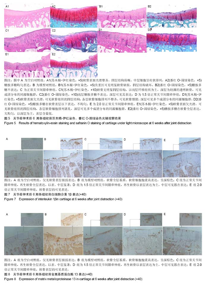
关节牵伸:是通过使用外固定架对关节进行牵伸,使关节达到去负荷的一项技术。关节牵伸技术应用于膝骨关节炎,目的是减轻关节软骨的机械应力,防止进一步的磨损和撕裂,进而刺激软骨细胞开启软骨组织修复;并且关节牵伸铰链允许活动,行走过程中关节液压力的变化可以改善软骨细胞营养,进一步促进软骨组织的修复。
基质金属蛋白酶:其家族成员具有相似的结构,一般由5个功能不同的结构域组成:疏水信号肽序列;前肽区,主要作用是保持酶原的稳定。当该区域被外源性酶切断后,基质金属蛋白酶原被激活;催化活性区,有锌离子结合位点,对酶催化作用的发挥至关重要;富含脯氨酸的铰链区;羧基末端区,与酶的底物特异性有关,其中酶催化活性区和前肽区具有高度保守性。各种基质金属蛋白酶间具有一定的底物特异性,但不是绝对的。同一种基质金属蛋白酶可降解多种细胞外基质成分,而某一种细胞外基质成分又可被多种基质金属蛋白酶降解,但不同酶的降解效率可不同。
文题释义:
关节牵伸:是通过使用外固定架对关节进行牵伸,使关节达到去负荷的一项技术。关节牵伸技术应用于膝骨关节炎,目的是减轻关节软骨的机械应力,防止进一步的磨损和撕裂,进而刺激软骨细胞开启软骨组织修复;并且关节牵伸铰链允许活动,行走过程中关节液压力的变化可以改善软骨细胞营养,进一步促进软骨组织的修复。
基质金属蛋白酶:其家族成员具有相似的结构,一般由5个功能不同的结构域组成:疏水信号肽序列;前肽区,主要作用是保持酶原的稳定。当该区域被外源性酶切断后,基质金属蛋白酶原被激活;催化活性区,有锌离子结合位点,对酶催化作用的发挥至关重要;富含脯氨酸的铰链区;羧基末端区,与酶的底物特异性有关,其中酶催化活性区和前肽区具有高度保守性。各种基质金属蛋白酶间具有一定的底物特异性,但不是绝对的。同一种基质金属蛋白酶可降解多种细胞外基质成分,而某一种细胞外基质成分又可被多种基质金属蛋白酶降解,但不同酶的降解效率可不同。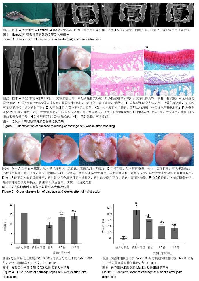
.jpg) 文题释义:
关节牵伸:是通过使用外固定架对关节进行牵伸,使关节达到去负荷的一项技术。关节牵伸技术应用于膝骨关节炎,目的是减轻关节软骨的机械应力,防止进一步的磨损和撕裂,进而刺激软骨细胞开启软骨组织修复;并且关节牵伸铰链允许活动,行走过程中关节液压力的变化可以改善软骨细胞营养,进一步促进软骨组织的修复。
基质金属蛋白酶:其家族成员具有相似的结构,一般由5个功能不同的结构域组成:疏水信号肽序列;前肽区,主要作用是保持酶原的稳定。当该区域被外源性酶切断后,基质金属蛋白酶原被激活;催化活性区,有锌离子结合位点,对酶催化作用的发挥至关重要;富含脯氨酸的铰链区;羧基末端区,与酶的底物特异性有关,其中酶催化活性区和前肽区具有高度保守性。各种基质金属蛋白酶间具有一定的底物特异性,但不是绝对的。同一种基质金属蛋白酶可降解多种细胞外基质成分,而某一种细胞外基质成分又可被多种基质金属蛋白酶降解,但不同酶的降解效率可不同。
文题释义:
关节牵伸:是通过使用外固定架对关节进行牵伸,使关节达到去负荷的一项技术。关节牵伸技术应用于膝骨关节炎,目的是减轻关节软骨的机械应力,防止进一步的磨损和撕裂,进而刺激软骨细胞开启软骨组织修复;并且关节牵伸铰链允许活动,行走过程中关节液压力的变化可以改善软骨细胞营养,进一步促进软骨组织的修复。
基质金属蛋白酶:其家族成员具有相似的结构,一般由5个功能不同的结构域组成:疏水信号肽序列;前肽区,主要作用是保持酶原的稳定。当该区域被外源性酶切断后,基质金属蛋白酶原被激活;催化活性区,有锌离子结合位点,对酶催化作用的发挥至关重要;富含脯氨酸的铰链区;羧基末端区,与酶的底物特异性有关,其中酶催化活性区和前肽区具有高度保守性。各种基质金属蛋白酶间具有一定的底物特异性,但不是绝对的。同一种基质金属蛋白酶可降解多种细胞外基质成分,而某一种细胞外基质成分又可被多种基质金属蛋白酶降解,但不同酶的降解效率可不同。