| [1] Stecco A,Stern R,Fantoni I,et al.Fascial Disorders: Implications for Treatment.PM R.2016; 8(2):161-168.[2] 常辉,李文惠.骨骼肌兴奋收缩偶联与细胞内的钙稳态[J].中国组织工程研究与临床康复,2010,14(50):9417-9420.[3] 马国震,李文惠,骆硕.骨骼肌三联管膜蛋白与兴奋收缩偶联[J].中国组织工程研究与临床康复,2011,15(2):347-350.[4] 张蓬,余跃,王世强.膜耦联的关键分子junctophilin.生理科学进展, 2009,40(3):209-213.[5] 刘琳,黄强民,汤莉.肌筋膜疼痛触发点[J].中国组织工程研究, 2014,18(46):7520-7527.[6] 刘琳,黄强民,刘庆广,等.肌筋膜触发点理论及其在运动康复临床实践中应用的研究进展[J].中国康复理论与实践,2016,22(10): 1167-1170.[7] Rossi D, Scarcella AM, Lorenzini S, et al. The Transmembrane Domain is Sufficient to Direct Junctophilin-1 Localization at the Junctional SR. Biophysical J.2016;10(3):98a. [8] Takeshima H, Hoshijima M, Song LS. Ca2+ microdomains organized by junctophilins. Cell Calcium.2015;(58):349-356. [9] Daniela Rossi, Angela Maria Scarcella, Stefania Lorenzini. et al.Localization of Junctophilin-1 at the Junctional Sarcoplasmic Reticulum Requires a Sequence in the Transmembrane Domain. Biophysical J.2017;112(3). [10] Sato D, Shannon TR, Bers DM. Sarcoplasmic Reticulum Structure and Functional Properties that Promote Long-Lasting Calcium Sparks. Biophysical J.2016;110(2):382-390. [11] Mustroph J,Wagemann O,Lebek S,et al.SR Ca2+-leak and disordered excitation-contraction coupling as the basis for arrhythmogenic and negative inotropic effects of acute ethanol exposure. J Mol Cell Cardiol.2018;116:81-90. [12] 蔡斌,戴尅戎.评估肌肉疲劳的中枢和外周起源[J].医用生物力学, 2015,30(2):192-196.[13] 李文惠,赵斌,闫万军.不同负荷间歇负重跑训练对老龄大鼠骨骼肌MG29蛋白的影响[J].体育科学,2010,30(9):73-81.[14] Shinichiro Yamamoto, Tetsuo Yamazaki, Shinji Komazaki. Contribution of calumin to embryo genesis through participation in the endoplasmic reticulum-associated degradation activity. Developmental Biology.2014; 393(1): 33-43. [15] 王海.不同信号调节肌纤维类型转换的研究[J].体育科技,2014, 35(5):74-75.[16] 武福云.Orai和STIM1相互作用机制研究[D].天津:南开大学, 2014.[17] Takeshima H, Komazaki S, NishiM, et al. Junctophilins: a novel family Of junctional membrane complex proteins. Mol Cell.2000;6:11-22. [18] Landstrom AP,Beavers DL,Wehrens XH.The junctophilin family of proteins: from bench to bedside. Trends Mol Med. 2014;20(6):353-362. [19] Komazaki S,Ito K,Takeshima H, et al.Deficiency of triad formation in developing skeletal muscle cells lacking junctophilin type 1.FEBS Lett.2002;524(1-3):225-229. [20] Hirata Y,Brotto M,Weisleder N,et al.Uncoupling Store-Operated Ca2+ Entry and Altered Ca2+ Release from Sarcoplasmic Reticulum through Silencing of Junctophilin Genes. Biophys J.2006;90(12):4418-4427. [21] 谢玉平,陈必义,王烈成.心肌肥厚或心肌衰竭中JUNCTOPH LIN-2和CAVEOLIN-3的表达变化[J].安徽医科大学学报,2011; 46(7):623-626.[22] Azad M,Khaledi N,Hedayati M.Effect of acute and chronic eccentric exercise on FOXO1 mRNA expression as fiber type transition factor in rat skeletal muscles. Gene.2016;584(2): 17-19. [23] Komazaki S,Nishi M,Takeshima H.Abnormal junctional membrane structures in cardiac myocytes expressing ectopic junctophilin type 1. Febs Letters.2003; 542(1-3):69-73. [24] 梁星光,吴博文,张维晨.Junctophilin 1在小鼠胚胎干细胞定向分化心肌细胞及大鼠胚胎心肌发生中的表征[J].浙江大学学报(医学版),2012,41(4):359-365.[25] 郭文静.JPH2敲减抑制小鼠心肌细胞SK2蛋白的表达[D].郑州:郑州大学,2014.[26] 李妙龄,欧贤红,李涛.U慢性心房颤动降低Junctophilin-2在心房中的表达研究[J].重庆医学,2016,45(22):3046-3048.[27] Reynolds JO,Quick AP,Wang Q,et al.Junctophilin-2 gene therapy rescues heart failure by normalizing RyR2-mediated Ca2+ release.Int J Cardiol.2016;225:371-380. [28] 潘宗富.Junctophilin 3和4在胚胎干细胞神经分化早期作用研究[D].浙江:浙江大学,2016.[29] Hai-Bo Zhang,Rong-Chang Li,Ming Xu,et al. Ultrastructural uncoupling between T-tubulesand sarco--plasmic reticulum in human heart failure, Cardiovasc. Res.2013; 98 (2):269-276. [30] Matsushita Y,Furukawa T,Kasanuki H, et al. Mutation of junctophilin type 2 associated with Hypertrophic cardiomyopathy. J HumanGenet.2007;52:543-548. [31] Landstrom AP, WeislederN, BataldenK B, et al. Mutations in JPH-2 encoded junctophilin-2 associated with hypertrophic cardiomyopathy in humans. J Mol Cell Cardiol. 2007;42: 1026-1035. [32] Landstrom AP,Kellen CA,Dixit SS,et al. Junctophilin-2 expression silencing causes cardiocyte hypertrophy and abnormal intracellular calcium-handling, Circ. Heart Fail. 2011;(4):214–223. [33] Zhang C,Chen B,Guo A,et al. Microtubule-mediated defects in junctophilin-2 traffick-ing contribute to myocyte transverse-tubule remodeling and Ca2+ handlingdysfunction in heart failure.Circulation.2014; (129):1742-1750. [34] Seixas AI,Holmes SE,Takeshima H, et al.Loss of junctophilin-3 con-tributes to Huntington disease-like 2 pathogenesis, Ann. Neurol.2012; (71):245–257. [35] Shannon H. Romer, Melissa Bautista, Daniel E. Hutcherson, et al. Architecture of Transverse Tubules and Triads in Huntington's Disease Skeletal Muscle. Biophysical J.2018; 114(3):469a. [36] Quick AP,Landstrom AP,Wang Q,et al. Novel Junctophilin-2 Mutation A405S Is Associated With Basal Septal Hypertrophy and Diastolic Dysfunction. JACC Basic Transl Sci. 2017;2(1): 56-67. [37] Quick AP,Landstrom AP,Wehrens XH. Novel JunJunctophilin-2 at the intersection of arrhythmia and pathologic cardiac remodeling. Heart Rhythm.2016;13(3): 753-754. [38] Roberts R.JPH2 Mutant Gene Causes Familial Hypertrophic Cardiomyopathy: A Possible Model to Unravel the Subtlety of Calcium-Regulated Contractility. JACC Basic Transl Sci. 2017; 2(1):68-70. [39] Ujihara Y, Mohri S, Katanosaka Y.Effects of induced Na+/Ca2+ exchanger overexpression on the spatial distribution of L-type Ca2+ channels and junctophilin-2 in pressure-overloaded hearts.Biochem Biophys Res Commun. 2016;480(4):564-569. |
.jpg)
.jpg)
.jpg) #br#
文题释义:#br#
兴奋-收缩偶联:是指将电兴奋过程和机械收缩联系起来的中介过程。其中介因子是钙离子,结构基础是三联管结构。过程:①肌膜动作电位沿横管传向肌细胞深处,并激活三联管上的L型钙通道;②L型钙通道的变构,钙离子的内流→激活终末池RYR→钙离子释放→胞质中钙离子浓度升高近百倍→与肌钙蛋白结合→肌肉收缩;③胞质内钙离子浓度升高的同时激活肌质网上的钙泵→回收钙离子→钙离子浓度降低→肌肉舒张。#br#
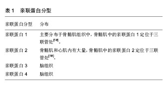
亲联蛋白功能:若亲联蛋白1和亲联蛋白2基因缺失,或干预RNA导致亲联蛋白1和亲联蛋白2基因抑制,均会导致骨骼肌三联管解偶联和心肌的二联管解偶联,打乱钙离子稳态,这说明亲联蛋白1和亲联蛋白2直接影响兴奋收缩偶联功能。亲联蛋白的主要功能是维持三联管的超微结构和构成偶联膜复合体,使细胞膜与肌浆网保持比较近的距离,从而为细胞膜到肌浆网之间的信号传导提供结构基础。
#br#
文题释义:#br#
兴奋-收缩偶联:是指将电兴奋过程和机械收缩联系起来的中介过程。其中介因子是钙离子,结构基础是三联管结构。过程:①肌膜动作电位沿横管传向肌细胞深处,并激活三联管上的L型钙通道;②L型钙通道的变构,钙离子的内流→激活终末池RYR→钙离子释放→胞质中钙离子浓度升高近百倍→与肌钙蛋白结合→肌肉收缩;③胞质内钙离子浓度升高的同时激活肌质网上的钙泵→回收钙离子→钙离子浓度降低→肌肉舒张。#br#
亲联蛋白功能:若亲联蛋白1和亲联蛋白2基因缺失,或干预RNA导致亲联蛋白1和亲联蛋白2基因抑制,均会导致骨骼肌三联管解偶联和心肌的二联管解偶联,打乱钙离子稳态,这说明亲联蛋白1和亲联蛋白2直接影响兴奋收缩偶联功能。亲联蛋白的主要功能是维持三联管的超微结构和构成偶联膜复合体,使细胞膜与肌浆网保持比较近的距离,从而为细胞膜到肌浆网之间的信号传导提供结构基础。