[1] KABEIL M, KAUVAR DS, BENNETT L, et al. Recent advances and the future of abdominopelvic and lower extremity vascular injury management. Semin Vasc Surg. 2023;36(2):268-282.
[2] SHOKOPLES BG, BERILLO O, COMEAU K, et al. P2RX7 gene knockout or antagonism reduces angiotensin II-induced hypertension, vascular injury and immune cell activation. J Hypertens. 2023;41(11):1701-1712.
[3] WANG X, ZHANG M, MAO C, et al. Icariin alleviates ferroptosis-related atherosclerosis by promoting autophagy in xo-LDL-induced vascular endothelial cell injury and atherosclerotic mice. Phytother Res. 2023;37(9): 3951-3963.
[4] TAI GJ, MA YJ, FENG JL, et al. NLRP3 inflammasome-mediated premature immunosenescence drives diabetic vascular aging dependent on the induction of perivascular adipose tissue dysfunction. Cardiovasc Res. 2025;121(1):77-96.
[5] PENG H, LV Y, LI C, et al. Cathepsin S inhibition in dendritic cells prevents Th17 cell differentiation in perivascular adipose tissues following vascular injury in diabetic rats. J Biochem Mol Toxicol. 2023;37(9):e23419.
[6] JIANG T, WEI Y, XU R, et al. Renal denervation alleviates vascular remodeling in spontaneously hypertensive rats by regulating perivascular adipose tissue. Hypertens Res. 2024;47(10):2760-2772.
[7] CHENG CK, DING H, JIANG M, et al. Perivascular adipose tissue: Fine-tuner of vascular redox status and inflammation. Redox Biol. 2023;62:102683.
[8] HILLOCK-WATLING C, GOTLIEB AI. The pathobiology of perivascular adipose tissue (PVAT), the fourth layer of the blood vessel wall. Cardiovasc Pathol. 2022;61:107459.
[9] PERSSON P, MARCHETTI M, FRIEDERICH-PERSSON M. Browning of perivascular adipose tissue prevents vascular dysfunction and reduces hypertension in angiotensin II-infused mice. Am J Physiol Regul Integr Comp Physiol. 2023;325(3):R290-R298.
[10] KATSIKI N, MIKHAILIDIS DP. Perivascular Adipose Tissue: Pathophysiological Links With Inflammation, Atherosclerosis, and Thrombosis. Angiology. 2022;73(3):195-196.
[11] KANWISCHER L, XU X, SAIFUDDIN AB, et al. Low levels of circulating methylated IRX3 are related to worse outcome after transcatheter aortic valve implantation in patients with severe aortic stenosis. Clin Epigenetics. 2023;15(1):149.
[12] LIU W, OH Y, YIN W, et al. The Combinatorial Role of Iroquois Homebinatorial Role of Iroquois Homeobox Genes 3 and 4 in the Compaction of the Ventricular Myocardium. Can J Cardiol. 2021;37(10, Supplement):S5.
[13] NAGEL S, MEYER C, POMMERENKE C. IRX-related homeobox gene MKX is a novel oncogene in acute myeloid leukemia. PLoS One. 2024;19(12): e0315196.
[14] MØLLER AL, VASAN RS, LEVY D, et al. Integrated omics analysis of coronary artery calcifications and myocardial infarction: the Framingham Heart Study. Sci Rep. 2023;13(1):21581.
[15] SMEMO S, TENA JJ, KIM KH, et al. Obesity-associated variants within FTO form long-range functional connections with IRX3. Nature. 2014; 507(7492):371-375.
[16] TU P, BHASIN S, HRUZ PW, et al. Genetic disruption of myostatin reduces the development of proatherogenic dyslipidemia and atherogenic lesions in Ldlr null mice. Diabetes. 2009;58(8):1739-1748.
[17] ZHOU X, CHEN Y, CUI L, et al. Advances in the pathogenesis of psoriasis: from keratinocyte perspective.Cell Death Dis. 2022;13(1):81.
[18] LI J, TIAN Z, ZHANG T, et al. Single-cell view and a novel protective macrophage subset in perivascular adipose tissue in T2DM. Cell Mol Biol Lett. 2024;29(1):148.
[19] BONDAREVA O, RODRÍGUEZ-AGUILERA JR, OLIVEIRA F, et al. Single-cell profiling of vascular endothelial cells reveals progressive organ-specific vulnerabilities during obesity. Nat Metab. 2022;4(11):1591-1610.
[20] TIMMONS JA, WENNMALM K, LARSSON O, et al. Myogenic gene expression signature establishes that brown and white adipocytes originate from distinct cell lineages. Proc Natl Acad Sci U S A. 2007; 104(11):4401-4406.
[21] RAO J, DJEFFAL Y, CHAL J, et al. Reconstructing human brown fat developmental trajectory in vitro. Dev Cell. 2023;58(21):2359-2375.
[22] CASTELLÁ M, BLASCO-ROSET A, PEYROU M, et al. Adipose tissue plasticity in pheochromocytoma patients suggests a role of the splicing machinery in human adipose browning. iScience. 2023;26(6):106847.
[23] LIU H, WANG X, GAO H, et al. Physiological and pathological characteristics of vascular endothelial injury in diabetes and the regulatory mechanism of autophagy. Front Endocrinol (Lausanne). 2023;14:1191426.
[24] TRAN KV, FITZGIBBONS T, MIN SY, et al. Distinct adipocyte progenitor cells are associated with regional phenotypes of perivascular aortic fat in mice. Mol Metab. 2018;9:199-206.
[25] HOU N, DU G, HAN F, et al. Irisin Regulates Heme Oxygenase-1/Adiponectin Axis in Perivascular Adipose Tissue and Improves Endothelial Dysfunction in Diet-Induced Obese Mice. Cell Physiol Biochem. 2017;42(2):603-614.
[26] GBD 2015 OBESITY COLLABORATORS, AFSHIN A, FOROUZANFAR MH, et al. Health Effects of Overweight and Obesity in 195 Countries over 25 Years. N Engl J Med. 2017;377(1):13-27.
[27] JIANG M, REN X, HAN L, et al. Associations between sarcopenic obesity and risk of cardiovascular disease: A population-based cohort study among middle-aged and older adults using the CHARLS. Clin Nutr. 2024;43(3):796-802.
[28] KOENEN M, HILL MA, COHEN P, et al. Obesity, Adipose Tissue and Vascular Dysfunction. Circ Res. 2021;128(7):951-968.
[29] TING P, WANG T, FU M, et al. Prevalence and inequalities of obesity and associated complications in China: A multicentre nationwide survey. Public Health. 2024;237:97-106.
[30] LI HF, LIU HT, CHEN PY, et al. Role of PVAT in obesity-related cardiovascular disease through the buffering activity of ATF3. iScience. 2022;25(12): 105631.
[31] HUANG CL, HUANG YN, YAO L, et al. Thoracic perivascular adipose tissue inhibits VSMC apoptosis and aortic aneurysm formation in mice via the secretome of browning adipocytes. Acta Pharmacol Sin. 2023;44(2):345-355.
[32] HAGBERG CE, SPALDING KL. White adipocyte dysfunction and obesity-associated pathologies in humans. Nat Rev Mol Cell Biol. 2024;25(4):333.
[33] PEIU SN, ZUGUN-ELOAE F, STOICA B, et al. Obesity-Induced PVAT Dysfunction and Atherosclerosis Development: The Role of GHSR-1a in Increased Macrophage Infiltration and Adipocytokine Secretion. J Cardiovasc Dev Dis. 2025;12(3):87.
[34] VAN DAM AD, BOON MR, BERBÉE JFP, et al. Targeting white, brown and perivascular adipose tissue in atherosclerosis development. Eur J Pharmacol. 2017;816:82-92.
[35] SHI H, GOO B, KIM D, et al. Perivascular adipose tissue promotes vascular dysfunction in murine lupus. Front Immunol. 2023;14:1095034.
[36] MU W, QIAN S, SONG Y, et al. BMP4-mediated browning of perivascular adipose tissue governs an anti-inflammatory program and prevents atherosclerosis. Redox Biol. 2021;43:101979.
[37] BROWN RM, WANG L, FU A, et al. Irx3 promotes gap junction communication between uterine stromal cells to regulate vascularization during embryo implantation. Biol Reprod. 2022;106(5):1000-1010.
[38] YAN XY, LUO YY, CHEN HJ, et al. IRX3 promotes adipose tissue browning and inhibits fibrosis in obesity-resistant mice. Int J Biochem Cell Biol. 2024; 175:106638.
[39] YAO J, WU D, ZHANG C, et al. Macrophage IRX3 promotes diet-induced obesity and metabolic inflammation. Nat Immunol. 2021;22(10):1268-1279.
[40] ZOU Y, LU P, SHI J, et al. IRX3 Promotes the Browning of White Adipocytes and Its Rare Variants are Associated with Human Obesity Risk. EBioMedicine. 2017;24:64-75.
[41] TU Q, LIU X, YAO X, et al. RETSAT associates with DDX39B to promote fork restarting and resistance to gemcitabine based chemotherapy in pancreatic ductal adenocarcinoma. J Exp Clin Cancer Res. 2022;41(1):274.
[42] JIANG X, HE Y, SHEN Q, et al. RETSAT Mutation Selected for Hypoxia Adaptation Inhibits Tumor Growth. Front Cell Dev Biol. 2021;9:744992.
[43] KIEFER MF, MENG Y, YANG N, et al. Intestinal retinol saturase is implicated in the development of obesity and epithelial homeostasis upon injury. Am J Physiol Endocrinol Metab. 2024;327(2):E203-E216.
[44] PANG XY, WANG S, JURCZAK MJ, et al. Retinol saturase modulates lipid metabolism and the production of reactive oxygen species. Arch Biochem Biophys. 2017;633:93-102.
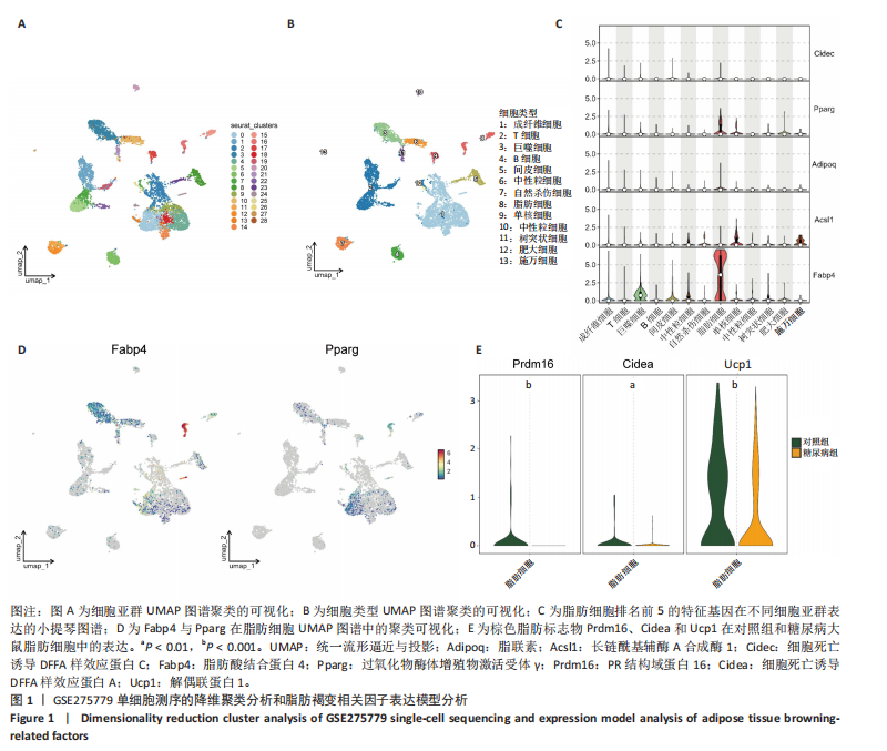
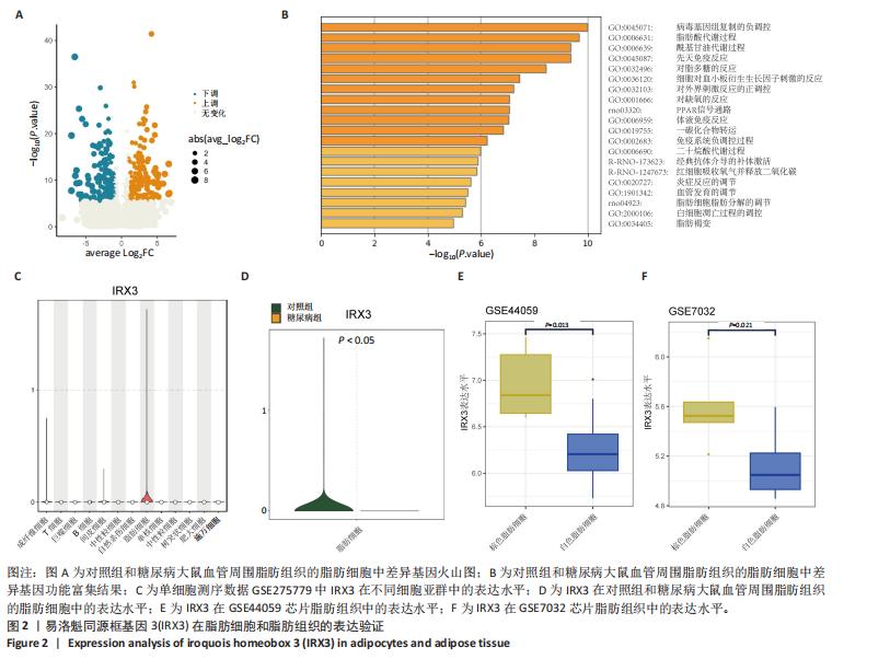
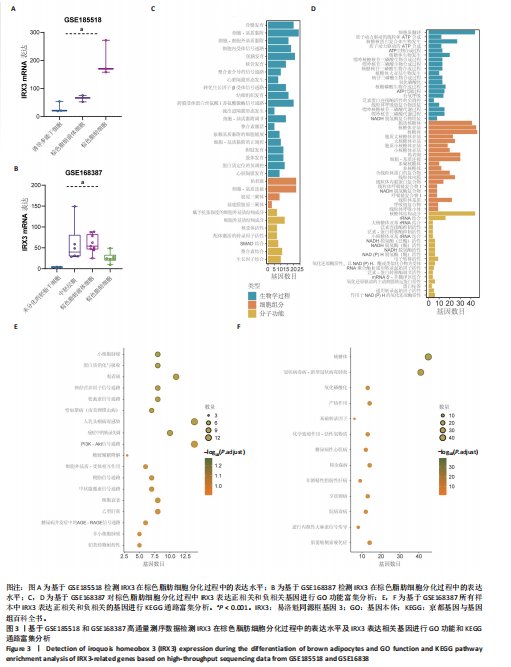
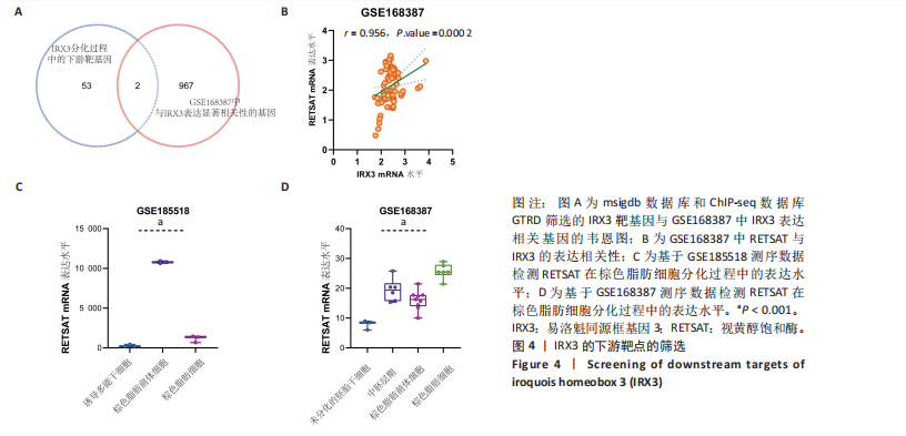
|