中国组织工程研究 ›› 2024, Vol. 28 ›› Issue (3): 360-365.doi: 10.12307/2023.838

• 组织工程口腔材料 tissue-engineered oral materials • 上一篇    下一篇

掺钕钇铝钙钛晶体激光结合两种再矿化制剂与早期釉质龋的再矿化

许颖华1,2,刘  婧2,尤  权2,文志豪2,高  璐2   

  1. 1上海市浦东新区眼病牙病防治所,上海市  200011;2大连医科大学口腔医学院,辽宁省大连市  116044
  • 收稿日期:2022-10-21 接受日期:2022-12-29 出版日期:2024-01-28 发布日期:2023-07-08
  • 通讯作者: 高璐,博士,副教授,硕士生导师,大连医科大学口腔医学院口腔解剖生理学教研室,辽宁省大连市 116044
  • 作者简介:许颖华,男,1980年生,上海市人,汉族,2007年大连医科大学毕业,硕士,副主任医师,主要从事口腔正畸学临床和基础研究。
  • 基金资助:
    浦东新区卫生和计划生育委员会卫生计生科研项目(PW2019A-21),项目负责人:许颖华;中国高等教育学会专项课题(2020SZYB23),项目负责人:高璐;辽宁省自然科学基金项目(2021-MS-293),项目负者人,高璐;辽宁省教育厅教改项目(2021-535),项目负者人:高璐

Effect of neodymium-doped:yttrium aluminum perovskite laser combined with two kinds of remineralizers on remineralization of early enamel caries

Xu Yinghua1, 2, Liu Jing2, You Quan2, Wen Zhihao2, Gao Lu2   

  1. 1Pudong New Area Eye Disease and Dental Disease Prevention & Treatment Center, Shanghai 200011, China; 2School of Stomatology, Dalian Medical University, Dalian 116044, Liaoning Province, China
  • Received:2022-10-21 Accepted:2022-12-29 Online:2024-01-28 Published:2023-07-08
  • Contact: Gao Lu, MD, Associated professor, Master’s supervisor, School of Stomatology, Dalian Medical University, Dalian 116044, Liaoning Province, China
  • About author:Xu Yinghua, Master, Associate chief physician, Pudong New Area Eye Disease and Dental Disease Prevention & Treatment Center, Shanghai 200011, China; School of Stomatology, Dalian Medical University, Dalian 116044, Liaoning Province, China
  • Supported by:
    Health and Family Planning Scientific Research Project of Pudong New Area, No. PW2019A-21 (to XYH); Special Project of China Association of Higher Education, No. 2020SZYB23 (to GL); the Natural Science Foundation of Liaoning Province, No. 2021-MS-293 (to GL); Education Reform Project of Department of Education in Liaoning Province, No. 2021-535 (to GL)

摘要:


文题释义:

掺钕钇铝钙钛晶体激光:波长为1 340 nm,水吸收率为2.700 832,激光的性能比较稳定,输出能量较高;与其他激光比较,组织穿透深度比较浅,临床上各项操作比较安全。在口腔医学临床应用方面,掺钕钇铝钙钛晶体激光可用于牙本质敏感、根管消毒、根尖封闭及种植体周围炎等,具有广阔的口腔医学临床应用前景。
早期牙釉质龋再矿化:牙体表面龋坏的发生和发展是一个动态变化的过程,主要表现为脱矿和再矿化的交替进行。早期牙釉质龋阶段在牙釉质表面脱矿形成白垩色斑块,此阶段如果采取相应治疗手段促进其再矿化可以抑制或者逆转龋病的进一步发展,这个过程称为早期牙釉质龋再矿化。


背景:近年来,多种激光被广泛应用于口腔医学相关的各种疾病,包括龋病的预防及治疗。

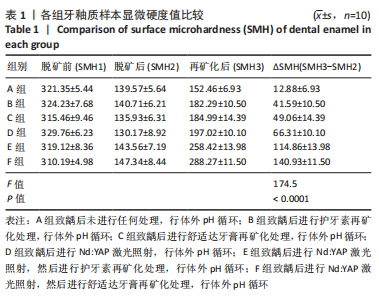
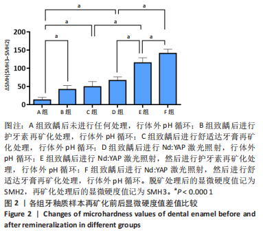
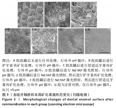
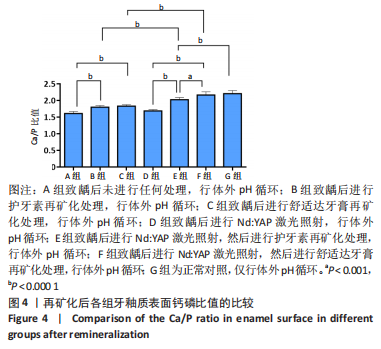
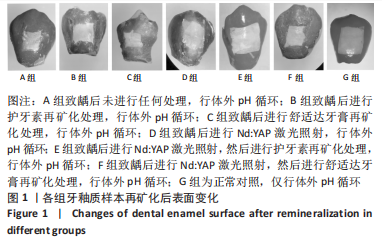
目的:探讨掺钕铝酸钇晶体(neodymium-doped: yttrium aluminum perovskite,Nd:YAP)激光联合两种再矿化制剂对早期釉质龋再矿化的作用。
方法:将60颗牙釉质块体外建立人工龋模型后,随机分为6组(n=10):A组不进行任何处理,行体外pH循环;B组进行护牙素(主要成分为酪蛋白磷酸钙复合体)再矿化处理,行体外pH循环;C组进行舒适达牙膏(主要成分为生物活性玻璃)再矿化处理,行体外pH循环;D组进行Nd:YAP激光照射,行体外pH循环;E组进行Nd:YAP激光照射,然后进行护牙素再矿化处理,行体外pH循环;F组进行Nd:YAP激光照射,然后舒适达牙膏再矿化处理,行体外pH循环;再矿化处理2次/d,实验周期20 d。G 组为正常对照,未致龋,未进行再矿化处理,仅行体外pH循环。实验结束后,检测各组牙釉质表面的显微硬度、形貌与Ca/P比值。

结果与结论:①B、C、D组牙釉质表面的硬度值高于A组(P < 0.000 1),E、F组牙釉质表面的硬度值明显高于B、C、D组(P < 0.000 1),F组牙釉质表面的硬度值明显高于E组(P < 0.000 1);②扫描电镜下可见,A组牙釉质表面存在大量的脱矿孔隙;B组牙釉质表面有矿物质沉积,沉积不均匀且较为疏松;C组牙釉质表面有大量矿物质沉积,且钙化团块间可见脱矿孔隙;D组牙釉质表面较为平整,脱矿孔隙较A组明显变小,釉柱间质有破坏;E组牙釉质表面矿物质沉积较厚,脱矿孔隙明显减少;F组牙釉质表面沉积的矿化物最为致密且均匀,脱矿孔隙细小;③B、C组牙釉质表面Ca/P比值明显高于A组(P < 0.000 1),E组牙釉质表面Ca/P比值明显高于B、C、D组(P < 0.000 1),F组牙釉质表面Ca/P比值高于E组(P < 0.001);③结果显示,生物活性玻璃、酪蛋白磷酸钙复合体、Nd:YAP激光在牙釉质脱矿后均具有促进牙釉质表面再矿化的作用,Nd:YAP激光联合生物活性玻璃或酪蛋白磷酸钙复合体能进一步加强牙釉质龋的再矿化作用,其中Nd:YAP激光和生物活性玻璃的联合使用效果最佳。

https://orcid.org/0000-0003-0698-5245(许颖华)

中国组织工程研究杂志出版内容重点:生物材料;骨生物材料口腔生物材料纳米材料缓释材料材料相容性组织工程

关键词: 牙釉质龋, 再矿化, 酪蛋白磷酸钙复合体, 生物活性玻璃, 掺钕钇铝钙钛晶体(Nd:YAP)激光, 显微硬度, 钙磷比, 联合治疗

Abstract: BACKGROUND: In recent years, a variety of lasers have been widely used in various diseases related to stomatology, including the prevention and treatment of dental caries. 
OBJECTIVE: To investigate the effect of neodymium-doped:yttrium aluminum perovskite (Nd:YAP) laser combined with two remineralizers on early enamel caries in vitro. 
METHODS: Early enamel caries models in vitro were artificially established by 60 enamel blocks and randomly divided into 6 groups (n=10). Group A did not undergo any treatment but underwent extracorporeal pH circulation. Group B underwent remineralization of dentin (the main component of casein phosphopeptide-amorphous calcium phosphate composite) and extracorporeal pH circulation. Group C underwent remineralization treatment of Sensodyne toothpaste (the main component of bioactive glass) and then underwent extracorporeal pH circulation. Group D received Nd:YAP laser irradiation and extracorporeal pH circulation. Group E was treated with Nd:YAP laser irradiation, with remineralization of dentin, and then with extracorporeal pH circulation. In group F, Nd:YAP laser irradiation was performed, and then Sensodyne toothpaste was used for remineralization, and the extracorporeal pH circulation was performed; the remineralization treatment was conducted twice a day, and the experimental period was 20 days. Group G was a normal control group, without caries or remineralization, but only underwent extracorporeal pH circulation. After the experiment, the microhardness, morphology and Ca/P ratio of the dental enamel surface were measured in each group.
RESULTS AND CONCLUSION: (1) The surface microhardness value of dental enamel in groups B, C and D was higher than that in group A (P < 0.000 1); the surface microhardness value of dental enamel in groups E and F was significantly higher than that in groups B, C and D (P < 0.000 1), and the surface microhardness value of dental enamel in group F was significantly higher than that in group E (P < 0.000 1). (2) Scanning electron microscopy showed that there were a lot of demineralized pores on the enamel surface of group A. There were mineral deposits on the enamel surface of group B, which were uneven and loose. In group C, there were a lot of mineral deposits on the enamel surface, and demineralized pores were found between the calcified masses. The enamel surface of group D was relatively flat; the demineralized pores were significantly smaller than that of group A, and the enamel column interstitium was damaged. In group E, the mineral deposits on the enamel surface were thicker and the demineralized pores were significantly reduced. The mineralized substances deposited on the enamel surface of group F were most dense and uniform and the demineralized pores were small. (3) The Ca/P ratio on the enamel surface of groups B and C was significantly higher than that of group A (P < 0.000 1); the Ca/P ratio on the enamel surface of group E was significantly higher than that of groups B, C and D (P < 0.000 1), and the Ca/P ratio on the enamel surface of group F was higher than that of group E (P < 0.001). (4) These findings indicate that bioactive glass, casein phosphopeptide-amorphous calcium phosphate composite, and Nd:YAP laser after enamel demineralization can promote the remineralization of early enamel caries. Nd:YAP laser combined with bioactive glass or casein phosphopeptide-amorphous calcium phosphate composite can further strengthen the remineralization of dental enamel caries, and the combination of Nd:YAP laser and bioactive glass has the best effect. 

Key words: enamel caries, remineralization, casein phosphopeptide-amorphous calcium phosphate composite, bioactive glass, neodymium-doped:yttrium aluminum perovskite laser, microhardness, calcium-phosphorus ratio, combination therapy

中图分类号: