[1] SAGHIRI MA, VAKHNOVETSKY J, VAKHNOVETSKY A, et al. Functional role of inorganic trace elements in dentin apatite tissue—Part 1: Mg, Sr, Zn, and Fe. J Trace Elem Med Biol. 2022;71:126932.
[2] JEONG J, KIM JH, SHIM JH, et al. Bioactive calcium phosphate materials and applications in bone regeneration. Biomater Res. 2019;23(1):4.
[3] Li XY, WEI L, LI JH, et al. Multifunctional SDF-1-loaded hydroxyapatite/polylactic acid membranes promote cell recruitment, immunomodulation, angiogenesis, and osteogenesis for biomimetic bone regeneration. Appl Mater Today. 2021;22:100942.
[4] YUAN B, CHEN H, ZHAO R, et al. Construction of a magnesium hydroxide/graphene oxide/hydroxyapatite composite coating on Mg–Ca–Zn–Ag alloy to inhibit bacterial infection and promote bone regeneration. Bioact Mater. 2022;18:354-367.
[5] CHIRICĂ IM, ENCIU AM, TITE T, et al. The physico-chemical properties and exploratory real-time cell analysis of hydroxyapatite nanopowders substituted with Ce, Mg, Sr, and Zn (0.5–5 at.%). Materials (Basel). 2021;14(14):3808.
[6] 代钊,汪大林.锶取代羟基磷灰石的制备方法和生物学特征[J].中国组织工程研究,2018,22(6):938-944.
[7] 罗进,严雅静,谭哲,等.羟基磷灰石抗菌改性的研究与应用[J].中国组织工程研究,2020,24(16):2606-2613.
[8] HASSAN AA, RADWAN HA, ABDELAAL SA, et al. Polycaprolactone based electrospun matrices loaded with Ag/hydroxyapatite as wound dressings: morphology, cell adhesion, and antibacterial activity. Int J Pharm. 2021;593:120143.
[9] 欧林结,潘育松,潘成岭.离子掺杂羟基磷灰石的抗菌性能研究进展[J].中国陶瓷,2021,57(11):8-14.
[10] 唐亚楠,高腾,任贵云.掺锌羟基磷灰石的制备及生物性能[J].中国组织工程研究,2022,26(16):2602-2607.
[11] 高建勇,田刚,朱强,等.锶掺杂改性羟基磷灰石的生物学性能研究[J].第二军医大学学报,2016,37(2):138-144.
[12] FRASNELLI M, CRISTOFARO F , SGLAVO VM, et al. Synthesis and characterization of strontium-substituted hydroxyapatite nanoparticles for bone regeneration. Mater Sci Eng C Mater Biol Appl. 2017;71:653-662.
[13] 晏金超.3D打印挤出沉积制备金属阳离子掺杂HA骨支架研究[D].南昌:南昌大学,2020.
[14] MOU P, PENG H, ZHOU L, et al. A novel composite scaffold of Cu-doped nano calcium-deficient hydroxyapatite/multi-(amino acid) copolymer for bone tissue regeneration. Int J Nanomed. 2019;14: 3331-3343.
[15] WEBSTER TJ, ERGUN C, DOREMUS RH, et al. Hydroxylapatite with substituted magnesium, zinc, cadmium, and yttrium. II. Mechanisms of osteoblast adhesion. J Biomed Mater Res. 2002;59(2):312-317.
[16] OHTSU N, KAKUCHI Y, OHTSUKI T. Antibacterial effect of zinc oxide/hydroxyapatite coatings prepared by chemical solution deposition. Appl Surf Sci. 2018;445:596-600.
[17] 贺琪,贡晶觉,杨蕾,等.不同载银量载银羟基磷灰石细胞相容性与成骨活性的对比研究[J].上海交通大学学报(医学版),2016, 36(12):1697-1701.
[18] HERKENDELL K, SHUKLA VR, PATEL AK, et al. Domination of volumetric toughening by silver nanoparticles over interfacial strengthening of carbon nanotubes in bactericidal hydroxyapatite biocomposite. Mater Sci Eng C Mater Biol Appl. 2014;34(1):455-467.
[19] 马晓雨,刘永佳,朱邦尚.镁掺杂纳米羟基磷灰石的制备及其在载药方面的应用[J].无机化学学报,2018,34(5):917-924.
[20] KOŁODZIEJSKA B, STĘPIEŃ N, KOLMAS J. The influence of strontium on bone tissue metabolism and its application in osteoporosis treatment. Int J Mol Sci. 2021;22(12):6564.
[21] TSAI SW, HSU YW, PAN WL, et al. The effect of strontium-substituted hydroxyapatite nanofibrous matrix on osteoblast proliferation and differentiation. Membranes (Basel). 2021;11(8):624.
[22] HU B, MENG ZD, ZHANG YQ, et al. Sr-HA scaffolds fabricated by SPS technology promote the repair of segmental bone defects. Tissue Cell. 2020;66:101386.
[23] LI K, LI SY, AI FR, et al. Fabrication and characterization of Sr-doped hydroxyapatite porous scaffold. JOM. 2021;73(6):1745-1753.
[24] QI YP, MAI S, YE Z, et al. Biomimetic fabrication and characterization of collagen/strontium hydroxyapatite nanocomposite. Mater Lett. 2020;274:127982.
[25] SAGHIRI MA, ASATOURIAN A, ORANGI J, et al. Functional role of inorganic trace elements in angiogenesis—Part II: Cr, Si, Zn, Cu, and S. Crit Rev Oncol Hematol. 2015;96(1):143-155.
[26] WU C, ZHOU Y, XU M, et al. Copper-containing mesoporous bioactive glass scaffolds with multifunctional properties of angiogenesis capacity, osteostimulation and antibacterial activity. Biomaterials. 2013;34(2): 422-433.
[27] ELRAYAH A, ZHI W, FENG S, et al. Preparation of micro/nano-structure copper-substituted hydroxyapatite scaffolds with improved angiogenesis capacity for bone regeneration. Materials (Basel). 2018; 11(9):1516.
[28] AI FR, CHEN LT, YAN JC, et al. Hydroxyapatite scaffolds containing copper for bone tissue engineering. J Sol-Gel Sci Technol. 2020;95(1):168-179.
[29] LV YR, CHEN YJ, ZHENG YP, et al. Evaluation of the antibacterial properties and in-vitro cell compatibilities of doped copper oxide/hydroxyapatite composites. Colloids Surf B Biointerfaces. 2022;209(Pt 2):112194.
[30] NARAYANAN V, SUMATHI S, NARAYANASAMY ANR. Tricomponent composite containing copper-hydroxyapatite/chitosan/polyvinyl pyrrolidone for bone tissue engineering. J Biomed Mater Res A. 2020; 108(9):1867-1880.
[31] PREDOI D, ICONARU SL, PREDOI MV, et al. Evaluation of antibacterial activity of zinc-doped hydroxyapatite colloids and dispersion stability using ultrasounds. Nanomaterials (Basel). 2019;9(4):515.
[32] DE LIMA CO, DE OLIVEIRA ALM, CHANTELLE L, et al. Zn-doped mesoporous hydroxyapatites and their antimicrobial properties. Colloids Surf B Biointerfaces. 2021;198:111471.
[33] LI JY, ZHU B, ZHANG YQ, et al. In vivo biological safety study of porous zinc oxide/hydroxyapatite composite materials. Zhongguo Xiu Fu Chong Jian Wai Ke Za Zhi. 2021;35(7):847-854.
[34] MALEKI-GHALEH H, SIADATI MH, OMIDI Y, et al. Synchrotron SAXS/WAXS and TEM studies of zinc doped natural hydroxyapatite nanoparticles and their evaluation on osteogenic differentiation of human mesenchymal stem cells. Mater Chem Phys. 2022;276:125346.
[35] MALEKI-GHALEH H, SIADATI MH, FALLAH A, et al. Antibacterial and cellular behaviors of novel zinc-doped hydroxyapatite/graphene nanocomposite for bone tissue engineering. Int J Mol Sci. 2021; 22(17):9564.
[36] MALEKI-GHALEH H, SIADATI HM, FALLAH A, et al. Effect of zinc-doped hydroxyapatite/graphene nanocomposite on the physicochemical properties and osteogenesis differentiation of 3D-printed polycaprolactone scaffolds for bone tissue engineering. Chem Eng J. 2021;426:13321.
[37] POON TKC, IYENGAR KP, JAIN VK. Silver nanoparticle (AgNP) technology applications in trauma and orthopaedics. J Clin Orthop Trauma. 2021; 21:101536.
[38] SAGHIRI MA, VAKHNOVETSKY J, VAKHNOVETSKY A, et al. Functional role of inorganic trace elements in dentin apatite tissue-part III: Se, F, Ag, and B. J Trace Elem Med Biol. 2022;72:126990.
[39] SEVENCAN A, KARTAL DOYUK E, KÖSE N. Silver ion doped hydroxyapatite-coated titanium pins prevent bacterial colonization. Jt Dis Relat Surg. 2021;32(1):35-41.
[40] BEE SL, BUSTAMI Y, UL-HAMID A, et al. Synthesis of silver nanoparticle-decorated hydroxyapatite nanocomposite with combined bioactivity and antibacterial properties. J Mater Sci Mater Med. 2021;32(9):106.
[41] YANG YW, CHENG Y, DENG F, et al. A bifunctional bone scaffold combines osteogenesis and antibacterial activity via in situ grown hydroxyapatite and silver nanoparticles. Bio-Des Manuf. 2021;4(3): 452-468.
[42] PATERSON TE, SHI R, TIAN JJ, et al. Electrospun scaffolds containing silver-doped hydroxyapatite with antimicrobial properties for applications in orthopedic and dental bone surgery. J Funct Biomater. 2020;11(3):58.
[43] CHOUBEY R, CHOUHAN R, BAJPAI AK, et al. Silver hydroxyapatite (AgHAP) reinforced nanocomposites of poly (methyl methacrylate)-poly (ɛ-caprolactone) as hybrid orthopedic materials. Int J Polym Mater. 2020;70(11):782-796.
[44] YOSHIZAWA S, BROWN A, BARCHOWSKY A, et al. Magnesium ion stimulation of bone marrow stromal cells enhances osteogenic activity, simulating the effect of magnesium alloy degradation. Acta Biomater. 2014;10(6):2834-2842.
[45] 史月华.掺镁羟基磷灰石涂层对种植体骨结合的影响[D].杭州:浙江大学,2014.
[46] ZHAO XN, YANG Z, LIU QY, et al. Potential load-bearing bone substitution material: carbon-fiber-reinforced magnesium-doped hydroxyapatite composites with excellent mechanical performance and tailored biological properties. ACS Biomater Sci Eng. 2022;8(2):921-938.
[47] WU ZQ, MENG ZL, WU QJ, et al. Biomimetic and osteogenic 3D silk fibroin composite scaffolds with nano MgO and mineralized hydroxyapatite for bone regeneration. J Tissue Eng. 2020;11: 2041731420967791.
[48] ANITA LETT J, SAGADEVAN S, LÉONARD E, et al. Bone tissue engineering potentials of 3D printed magnesium-hydroxyapatite in polylactic acid composite scaffolds. Artif Organs. 2021;45(12):1501-1512.
[49] ULLAH I, ZHANG WC, YANG L, et al. Impact of structural features of Sr/Fe co-doped HAp on the osteoblast proliferation and osteogenic different-iation for its application as a bone substitute. Mater Sci Eng C Mater Biol Appl. 2020;110:110633.
[50] YANG L, ULLAH I, YU K, et al. Bioactive Sr2+/Fe3+co-substituted hydro-xyapatite in cryogenically 3D printed porous scaffolds for bone tissue engineering. Biofabrication. 2021;13(3):84.
[51] YANG L, JIN SY, SHI L, et al. Cryogenically 3D printed biomimetic scaffolds containing decellularized small intestinal submucosa and Sr2+/Fe3+ co-substituted hydroxyapatite for bone tissue engineering. Chem Eng J. 2022;431(P4):133459.
[52] ULLAH I, SIDDIQUI MA, LIU H, et al. Mechanical, biological, and antibacterial characteristics of plasma-sprayed (Sr, Zn) substituted hydroxyapatite coating. ACS Biomater Sci Eng. 2020;6(3):1355-1366.
[53] ZHONG ZY, WU XD, WANG YF, et al. Zn/Sr dual ions-collagen co-assembly hydroxyapatite enhances bone regeneration through procedural osteo-immunomodulation and osteogenesis. Bioact Mater. 2022;10:195-206.
[54] HASSAN M, SULAIMAN M, YUVARAJU PD, et al. Biomimetic PLGA/strontium-zinc nano hydroxyapatite composite scaffolds for bone regeneration. J Funct Biomater. 2022;13(1):13.
[55] LIU XJ, MA YH, CHEN MJ, et al. Ba/Mg co-doped hydroxyapatite/PLGA composites enhance X-ray imaging and bone defect regeneration. J Mater Chem B. 2021;9(33):6691-6702.
[56] YAN TT, JIANG ZM, LI P, et al. Novel hydroxyapatite whiskers modified by silver ion and nano zinc oxide used for bone defect repairment. Coatings. 2021;11(8):957.
[57] SINULINGGA K, SIRAIT M, SIREGAR N, et al. Investigation of antibacterial activity and cell viability of Ag/Mg and Ag/Zn Co-doped hydroxyapatite derived from natural limestone. ACS Omega. 2021;6(49):34185-34191.
[58] BENSALEM A, KUCUKOSMAN OK, RASZKIEWICZ J, et al. Synthesis, characterization, bactericidal activity, and mechanical properties of hydroxyapatite nano powders impregnated with silver and zinc oxide nanoparticles (Ag-ZnO-Hap). Ceram Int. 2021;47(15):21319-21324.
[59] MARLIANA BIY, SYAZWAN MNM, AHMAD-FAUZI MN, et al. Influence of ternary divalent cations (Mg2+, Co2+, Sr2+) substitution on the physicochemical, mechanical and biological properties of carbonated hydroxyapatite scaffolds. J Aust Ceram Soc. 2021;57(5):1499-1510.
[60] XIAO SJ, WANG M, WANG LP, et al. Environment-friendly synthesis of trace element Zn, Sr, and F codoping hydroxyapatite with non-cytotoxicity and improved osteoblast proliferation and differentiation. Biol Trace Elem Res. 2018;185(1):148-161.
[61] SPRIO S, DAPPORTO M, PRETI L, et al. Enhancement of the biological and mechanical performances of sintered hydroxyapatite by multiple ions doping. Front Mater. 2020;7:224. |
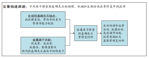

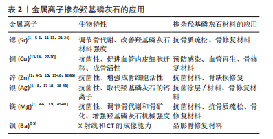
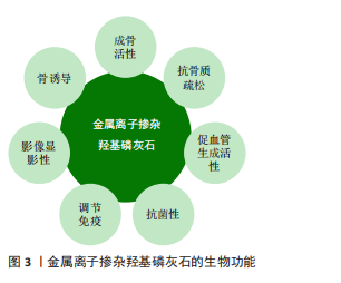 离子掺杂后的羟基磷灰石具有良好的相容性,亦可与聚合物、无机物进一步复合形成新材料用于骨修复。近年来有关金属离子掺杂改性羟基磷灰石骨修复材料的研究取得了一定的进展,见表2,但目前的研究多关注一种离子掺杂羟基磷灰石的制备、形态结构的变化或离子掺杂羟基磷灰石某一性能的研究;缺乏对常见离子改性后羟基磷灰石的形态学、结构学、生物相容性、成骨成血管活性及抗菌性等性能的系统研究;各离子具有一定的毒性,低浓度掺杂的羟基磷灰石具有良好的生物相容性,但高浓度掺杂时则会出现毒副反应,有关各离子掺杂的最佳浓度缺乏统一标准。
 该综述就离子掺杂羟基磷灰石的制备方法、常见功能离子一元和多元掺杂羟基磷灰石后的生物相容性、毒性、机械性能、成骨活性、成血管活性及抗菌性等特性进行了系统总结。同时,文章存在一定局限性,未对离子掺杂后羟基磷灰石的结晶学参数和形态参数进行阐述;未对各离子改性后羟基磷灰石的性能进行横向对比;未对各离子参与骨代谢的机制进行详细讨论;对掺杂后羟基磷灰石的抗菌、成血管及免疫调节机制未进行深入阐释。