中国组织工程研究 ›› 2022, Vol. 26 ›› Issue (12): 1861-1865.doi: 10.12307/2022.508

• 骨科植入物 orthopedic implant • 上一篇    下一篇

计算机导航下设计经皮穿刺置入空心椎弓根螺钉的轨道

袁翠华1,林  旺1,蒋  垚2,刘成招1   

  1. 1福建医科大学附属闽东医院骨科,福建省福安市   355000;2苏州大学医学部临床医学院,江苏省苏州市   215000
  • 收稿日期:2021-03-09 修回日期:2021-03-10 接受日期:2021-06-09 出版日期:2022-04-28 发布日期:2021-12-14
  • 通讯作者: 林旺,硕士,副主任医师,福建医科大学附属闽东医院骨科,福建省福安市 355000 蒋垚,硕士,主任医师,博士研究生导师,教授,苏州大学医学部临床医学院,江苏省苏州市 215000
  • 作者简介:袁翠华,男,1979 年生,汉族,2016年苏州大学毕业,博士,副主任医师,主要从事脊柱外科方面的研究。
  • 基金资助:
    福建省医学创新课题(2020CXA059),项目负责人:袁翠华;福建省自然基金卫生行业联合资金面上项目(2017J01395),项目负责人:袁翠华

Screw track in computer navigation-assisted percutaneous hollow pedicle screw placement

Yuan Cuihua1, Lin Wang1, Jiang Yao2, Liu Chengzhao1    

  1. 1Department of Orthopedics, Mindong Hospital Affiliated to Fujian Medical University, Fuan 355000, Fujian Province, China; 2School of Clinical Medicine, Medical College of  Soochow University, Suzhou 215000, Jiangsu Province, China
  • Received:2021-03-09 Revised:2021-03-10 Accepted:2021-06-09 Online:2022-04-28 Published:2021-12-14
  • Contact: Lin Wang, Master, Associate chief physician, Department of Orthopedics, Mindong Hospital Affiliated to Fujian Medical University, Fuan 355000, Fujian Province, China Jiang Yao, Master, Chief physician, Doctoral supervisor, Professor, School of Clinical Medicine, Medical College of Soochow University, Suzhou 215000, Jiangsu Province, China
  • About author:Yuan Cuihua, MD, Associate chief physician, Department of Orthopedics, Mindong Hospital Affiliated to Fujian Medical University, Fuan 355000, Fujian Province, China
  • Supported by:
    Fujian Medicine Innovation Project, No. 2020CXA059 (to YCH); Natural Health Industry Joint Fund Project, No. 2017J01395 (to YCH)

摘要:

文题释义:
二维计算机导航:应用二维计算机导航系统通过X射线即时获得透视图,然后通过计算机导航系统辅助引导下置钉,无需术前采集图像,而且术中可实时显示图像,但不足之处在于所得图像为二维图像,不如三维图像精确。
依赖CT三维导航:术前对目标椎体进行薄层CT扫描三维重建,制定手术方案,术中在三维CT导航引导下放置椎弓根钉。由于CT良好的三维图像,可实时显像,置钉具有较高的精确性。

背景:经皮穿刺安放椎弓根钉时,不同的术者对进钉点及进钉角度各家报道不一,使得经皮微创椎弓根螺钉技术的应用受到了限制。目前行经皮椎弓根钉技术治疗胸腰椎骨折时,大多根据术中经验或依据术中X射线反复透视引导下置钉,患者和术者受到医源性损害较大。
目的:分别从尸体解剖学和影像学角度研究计算机导航下设计经皮穿刺置入椎弓根螺钉轨道的可行性。
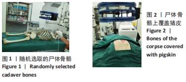
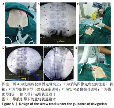
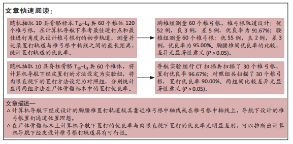
方法:①随机抽取10具骨骼标本T10-L3共60个椎体120个椎弓根,在计算机导航下参考最佳进钉点和最佳进钉角度来设计椎弓根钉的初步轨道,测量并记录置钉轨道与椎弓根中轴线之间的最长距离,统计置钉轨道的优良率,分析导航下置钉轨道的精确性;②随机抽取10具脊柱骨骼T10-L3共60个椎体,将计算机导航下经皮置钉的方法设定为实验组,将肉眼直视下的置钉方法设定为对照组,分别统计应用两组方法在尸体骨骼标本中的置钉优良率。
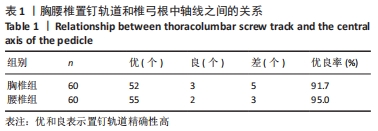
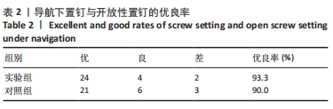
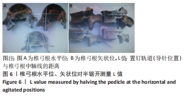
结果与结论:①胸椎组测量60个椎弓根,椎弓根轨道设计:优52例,良3例,差5例,优良率为91.67%;腰椎组测量60个椎弓根:优55例,良2例,差3例,优良率为95.00%;胸腰椎间优良率的比较,差异无显著性意义(P > 0.05);②导航实验组行CT扫描共扫描了30个椎弓根,置钉优良率96.67%;对照组共扫描了30个椎弓根,置钉优良率90.00%,两组间比较差异无显著性意义(P > 0.05);③计算机导航下经皮设计的胸腰椎置钉轨道极其靠近椎弓根中轴线或在椎弓根中轴线上,导航下设计的椎弓根置钉通道位置理想;在尸体骨骼标本上计算机导航下置钉的优良率与肉眼直视下置钉的优良率无明显差别,可以推断出计算机导航下经皮设计椎弓根钉轨道具有可行性。

https://orcid.org/0000-0002-5130-4069 (袁翠华) 

中国组织工程研究杂志出版内容重点:人工关节;骨植入物;脊柱;骨折;内固定;数字化骨科;组织工程

关键词: 计算机, 导航, 解剖, 微创, 椎弓根螺钉

Abstract: BACKGROUND: Percutaneous minimally invasive pedicle screw placement has limited the use of minimally invasive percutaneous pedicle screw technology, resulting in clinical confusion caused by different reports on the insertion point and angle of the pedicle screw. At present, when percutaneous pedicle screw technique is used in the treatment of thoracolumbar fracture, most of the screws are placed according to experience or the guidance of intraoperative X-ray repeated fluoroscopy. Thus, the patients and the operators suffer great iatrogenic damage.  
OBJECTIVE: To study the feasibility of percutaneous pedicle screw orbit design respectively from the angle of body anatomy and imaging under the navigation design of precision of the orbit.
METHODS:  (1) Ten bone specimens (T10-L3, a total of 120 vertebral pedicles) were randomly selected, and the preliminary track for pedicle screw was designed based on the best entry points and the best needling angle under computer navigation. The maximum distance from pedicle screw track to the central axis of the vertebral pedicle was measured and recorded. The excellent and good rate of screw placement was counted to analyze the accuracy of the screw track under navigation. (2) Ten spinal bones (T10-L3, a total of 60 vertebral pedicles) were randomly selected. Cases treated with computer navigation-assisted percutaneous pedicle screw placement were set as experimental group. Meanwhile, cases treated with screw placement method under direct vision with naked eyes were set as control group. The excellent and good rate of screw placement among the bone specimens of corpses in both groups was counted and analyzed, separately.  
RESULTS AND CONCLUSION: (1) The measurement of 60 vertebral pedicles in the thoracic spine group and the design of pedicle screw track: There were 52 excellent cases, 3 good cases, and 5 poor cases, with an excellent and good rate of 91.67%. The measurement of 60 vertebral pedicles in the lumbar spine group: There were 55 excellent cases, 2 good cases, and 3 poor cases, with an excellent and good rate of 95.00%. No significant difference in the excellent and good rate was detected between thoracic and lumbar spines (P > 0.05). (2) CT scanning was carried out on a total of 30 vertebral pedicles in the navigation experimental group, with an excellent and good rate of screw placement of 96.67%. A total of 30 vertebral pedicles were scanned in the control group, with an excellent and good rate of screw placement of 90.00%. No significant difference in the excellent and good rate was detected between these two groups (P > 0.05). (3) The screw track of computer navigation-assisted percutaneous pedicle screw placement for thoracic and lumbar spines is extremely close to the central axis of the vertebral pedicle or located at the central axis of the vertebral pedicle, and the pedicle screw track designed under navigation is ideal. The excellent and good rate of screw placement among the bone specimens of corpses shows no apparent difference under navigation and direct vision with naked eyes. Therefore, it can be inferred that the pedicle screw track designed with computer navigation-assisted percutaneous placement is feasible.

Key words: computer, navigation, anatomy, minimally invasive, pedicle screw

中图分类号: