[1] ALSHAMI AM. Prevalence of spinal disorders and their relationships with age and gender. Saudi Med J. 2015;36(6):725-730.
[2] KOLENKIEWICZ M, WŁODARCZYK A, WOJTKIEWICZ J. Diagnosis and Incidence of Spondylosis and Cervical Disc Disorders in the University Clinical Hospital in Olsztyn, in Years 2011–2015. BioMed Res Int. 2018; 2018:5643839.
[3] ABUMI K, ITOH H, TANEICHI H, et al. Transpedicular Screw Fixation for Traumatic Lesions of the Middle and Lower Cervical Spine. J Spinal Disord. 1994;7(1):19-28.
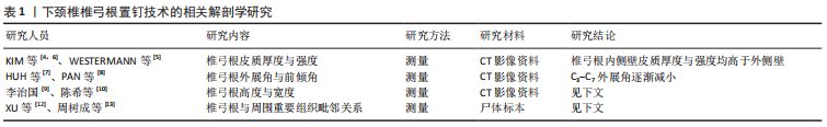
[4] KIM M, CHO H, KWAK D. A new anatomical approach of cervical lateral mass for cervical pedicle screw and paravertebral foramen screw insertion. PLOS ONE. 2019;14(7):e0219119.
[5] WESTERMANN L, SPEMES C, EYSEL P, et al. Computer tomography-based morphometric analysis of the cervical spine pedicles C3-C7. Acta Neurochir (Wien). 2018;160(4):863-871.
[6] KIM M, CHO H, KWAK D, et al. Characteristics of regional bone quality in cervical vertebrae considering BMD: Determining a safe trajectory for cervical pedicle screw fixation. J Orthop Res. 2018;36(1):217-223.
[7] HUH J, HYUN JH, PARK HG, et al. Three Dimensional Measurement of Ideal Trajectory of Pedicle Screws of Subaxial Cervical Spine Using the Algorithm Could Be Applied for Robotic Screw Insertion. J Korean Neurosurg Soc. 2019;62(4):376-381.
[8] PAN Z, ZHONG J, XIE S, et al. Accuracy and Safety of Lateral Vertebral Notch-Referred Technique Used in Subaxial Cervical Pedicle Screw Placement. Oper Neurosurg (Hagerstown). 2019;17(1):52-60.
[9] 李治国. 颈椎椎弓根形态个体化术前应用CT测量评价的临床意义[J]. 中国CT和MRI杂志,2016,14(6):130-132.
[10] 陈希,刘新宇,杨青,等. 颈椎椎间孔螺钉、椎弓根螺钉与侧块螺钉钉道的影像学测量比较[J]. 中华骨科杂志,2020,40(19):1337-1347.
[11] ZHOU Z, WEN C, SUN Q, et al. Morphometric measurement of the cervical spine for minimally invasive pedicle screw fixation using reverse engineering and three-dimensional reconstruction. Int J Med Robot. 2017;13(3):e1765.
[12] Xu R,Kang A,ebraHein NA, et al. Anatomic Relation Between the Cervical Pedicle and the Adjacent Neural Structures. Spine. 1999;24(5): 451-454.
[13] 周树成,张鹏.下颈椎椎弓根相邻解剖结构的应用研究[J]. 局解手术学杂志,2016,25(1):30-32.
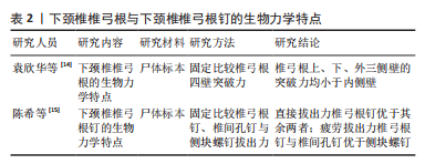
[14] 袁欣华,庞清江,许柯,等.下颈椎椎弓根通道穿透力及突破力的生物力学研究[J].现代实用医学,2012,24(10):1096-1098+1201.
[15] 陈希,刘新宇,杨青,等. 颈椎椎间孔螺钉、侧块螺钉及椎弓根螺钉的生物力学强度比较[J]. 中华骨科杂志,2020,40(4):236-243.
[16] 薛晓北,王玉冰,纪经涛,等.区域法下颈椎椎弓根钉植入技术的临床应用[J].中国修复重建外科杂志,2020,34(12):1515-1520.
[17] WU C, HUANG Z, PAN Z, et al. Coronal Multiplane Reconstructed Computed Tomography Image Determining Lateral Vertebral Notch-Referred Pedicle Screw Entry Point in Subaxial Cervical Spine: A Preclinical Study. World Neurosurg. 2017;103:322-329.
[18] LUO J, WU C, HUANG Z, et al. The accuracy of the lateral vertebral notch-referred pedicle screw insertion technique in subaxial cervical spine: a human cadaver study. Arch Orthop Trauma Surg. 2017;137(4):517-522.
[19] LIU J, LI Y, WU Y, et al. A novel method of cervical pedicle screw placement from C3 to C5 and its clinical applications. Spine (Phila Pa 1976). 2013;38(8):504-512.
[20] LI Y, LIU J, LIU Y, et al. Cervical pedicle screw fixation at C6 and C7 A cadaveric study. Indian J Orthop. 2015;49(4):465-470.
[21] 邹云龙,刘宇龙,李野,等. 颈椎椎弓根置钉精确度的临床研究[J]. 中华骨科杂志,2017,37(4):226-235.
[22] LEE JH, CHOI BK, HAN IH, et al. Cervical Pedicle Screw Placement Using Medial Funnel Technique. Korean J Spine. 2017;14(3):84-88.
[23] KARAIKOVIC EE, YINGSAKMONGKOL W, GAINES RW, et al. Accuracy of Cervical Pedicle Screw Placement Using the Funnel Technique. Spine. 2001;26(22):2456-2462.
[24] LIU B, LIU X, SHEN X, et al. The “slide technique”-a novel free-hand method of subaxial cervical pedicle screw placement. BMC Musculoskelet Disord. 2020;21(1):399.
[25] LEE S, SEO J, LEE MK, et al. Widening of the safe trajectory range during subaxial cervical pedicle screw placement: advantages of a curved pedicle probe and laterally located starting point without creating a funnel-shaped hole. J Neurosurg Spine. 2017;27(2):150-157.
[26] BURCEV A, PAVLOVA O, DIACHKOV K, et al. Easy method to simplify “freehand” subaxial cervical pedicle screw insertion. J Craniovertebr Junction Spine. 2017;8(4):390-395.
[27] PARK J, LEE JY, LEE BH, et al. Free-Hand Cervical Pedicle Screw Placement by Using Para-articular Minilaminotomy: Its Feasibility and Novice Neurosurgeons’ Experience. Global Spine J. 2020: 2192568220919089.
[28] 黄晓东,袁登荣,方弘伟,等. 经肌间隙入路椎弓根螺钉置入治疗下颈椎骨折脱位15例效果分析[J]. 中国乡村医药,2016,23(8):20-21.
[29] 方弘伟,袁登荣,黄晓东,等. 经肌间隙入路行下颈椎椎弓根螺钉置入的应用解剖与初步临床应用[J]. 中华创伤杂志,2015,31(8):699-703.
[30] 余伟,王蕾,何思峰,等. X射线透视辅助徒手法与CT三维图像导航下颈椎椎弓根置钉准确率的对比[J]. 中国组织工程研究,2017, 21(11):1758-1763.
[31] 刘亚军,乐晓峰,郑山,等. 计算机导航辅助颈椎椎弓根螺钉内固定技术[J]. 骨科临床与研究杂志,2018,3(3):182-187.
[32] 姜廷华,李继斌,王自鸿,等. CT导航置钉与传统X线透视徒手置钉法在下颈椎椎弓根置钉的临床对比[J]. 颈腰痛杂志,2018,39(4): 397-400.
[33] SHIMOKAWA N, TAKAMI T. Surgical safety of cervical pedicle screw placement with computer navigation system. Neurosurg Rev. 2017; 40(2):251-258.
[34] 黄圣斌,谢兆林,谭海涛,等. O型臂导航系统辅助颈椎椎弓根置钉的应用效果研究[J]. 广西医科大学学报,2018,35(7):976-979.
[35] SOURABH C, RAHMATULLAH BARH, LIM LW, et al. Cervical pedicle screw instrumentation is more reliable with O-arm-based 3D navigation: analysis of cervical pedicle screw placement accuracy with O-arm-based 3D navigation. Eur Spine J. 2018;27(11):2729-2736.
[36] WADA K, TAMAKI R, INOUE T, et al. Cervical Pedicle Screw Insertion Using O-Arm-Based 3D Navigation: Technical Advancement to Improve Accuracy of Screws. World Neurosurg. 2020;139:e182-e188.
[37] 毛克政,王庆德,梅伟,等. 3D打印个体化导板辅助颈椎椎弓根螺钉置钉的可行性研究[J]. 中华创伤杂志,2016,32(1):47-50.
[38] YU Z, ZHANG G, CHEN X, et al. Application of a novel 3D drill template for cervical pedicle screw tunnel design: a cadaveric study. Eur Spine J. 2017;26(9):2348-2356.
[39] 郝申申,刘志斌,王飞,等. 3D打印个性化导航模板在后路下颈椎椎弓根螺钉置入中的应用价值[J]. 中国组织工程研究,2018,22(11): 1701-1706.
[40] SUGAWARA T, KANEYAMA S, HIGASHIYAMA N, et al. Prospective Multicenter Study of a Multistep Screw Insertion Technique Using Patient-Specific Screw Guide Templates for the Cervical and Thoracic Spine. Spine. 2018;43(23):1685-1694.
[41] 姜良海,谭明生,杨峰,等. 标杆型3D打印导板辅助颈椎椎弓根置钉的临床应用[J]. 中华骨科杂志,2016,36(5):257-264.
[42] 张东升,王建华,李洪吉,等. 改良金属个性化导航模板辅助颈椎椎弓根置钉的准确性[J]. 脊柱外科杂志,2019,17(5):340-344.
[43] LIEBERMAN IH, KISINDE S, HESSELBACHER S. Robotic-Assisted Pedicle Screw Placement During Spine Surgery. JBJS Essent Surg Tech. 2020; 10(2):e0020.
[44] 吕振东,陈智,韩应超,等. 机器人辅助颈椎后路椎弓根螺钉置入手术治疗颈椎病的置钉精确度与临床疗效[J]. 骨科临床与研究杂志,2020,5(3):131-137.
[45] SU X, LV Z, CHEN Z, et al. Comparison of Accuracy and Clinical Outcomes of Robot-Assisted Versus Fluoroscopy-Guided Pedicle Screw Placement in Posterior Cervical Surgery. Global Spine J. 2020:2192568220960406.
[46] ZHU S, ZHAO Z, PAN Y, et al. Markerless robotic pedicle screw placement based on structured light tracking. Int J Comput Assist Radiol Surg. 2020;15(8):1347-1358.
[47] 李绪辉. 后路微创经皮椎弓根钉内固定术治疗下颈椎骨折脱位的效果观察[J]. 广东医科大学学报,2020,38(4):417-420.
[48] 郎昭,田伟,袁强,等. 术中即时三维导航引导经皮微创椎弓根螺钉内固定治疗颈椎骨折的临床研究[J]. 中华外科杂志,2015,53(10): 752-756.
[49] KOMATSUBARA T, TOKIOKA T, SUGIMOTO Y, et al. Minimally Invasive Cervical Pedicle Screw Fixation by a Posterolateral Approach for Acute Cervical Injury. Clin Spine Surg. 2017;30(10):466-469.
[50] 唐文,邱兴庭,赵凯. 基于CT影像的下颈椎椎弓根钉道虚拟构建法[J]. 中华骨科杂志,2020,40(20):1409-1419.
[51] 赖必华,吴建斌,叶宏,等. 导向器结合钉道内壁探查法在下颈椎椎弓根螺钉置入中应用研究[J]. 中国骨伤,2017,30(9):805-809.
[52] WANG Q, XING R, ZENG Y. Design and application of subaxial cervical pedicle screw placement guide device. Exp Ther Med. 2019;17(6): 4357-4362. |