[1] KALLER P, HOHENBERGER G, HOLWEG P, et al. Fußblock [Ankle block]. Oper Orthop Traumatol. 2020;32(1):29-34.
[2] MEDINA MCKEON JM, HOCH MC. The Ankle-Joint Complex: A Kinesiologic Approach to Lateral Ankle Sprains. J Athl Train. 2019;54(6):589-602.
[3] SONG K, WIKSTROM EA. Plausible mechanisms of and techniques to assess ankle joint degeneration following lateral ankle sprains: a narrative review. Phys Sportsmed. 2019;47(3):275-283.
[4] AIYER AA, ZACHWIEJA EC, LAWRIE CM, et al. Management of Isolated Lateral Malleolus Fractures. J Am Acad Orthop Surg. 2019;27(2):50-59.
[5] IELAPI A, FORWARD M, DE BEULE M. Computational and experimental evaluation of the mechanical properties of ankle foot orthoses: A literature review. Prosthet Orthot Int. 2019;43(3):339-348.
[6] 张峻霞,窦树斐,苏海龙,等.上、下楼梯步态参数变化特征研究[J].医用生物力学,2016,31(3):266-271.
[7] PARK S, LEE S, YOON J, et al. Finite element analysis of knee and ankle joint during gait based on motion analysis. Med Eng Phys. 2019;63:33-41.
[8] 宫玉锁,周君,李盛华,等.骨科三维有限元研究进展[J].世界最新医学信息文摘,2019,19(93):40-41+43.
[9] 孙卫东,温建民.足部有限元建模方法应用现状[J]. 中国组织工程研究与临床康复,2010,14(13):2457-2461.
[10] WANG CW, MUHEREMU A, BAI JP. Use of three-dimensional finite element models of the lateral ankle ligaments to evaluate three surgical techniques. J Int Med Res. 2018;46(2):699-709.
[11] MONDAL S, GHOSH R. A numerical study on stress distribution across the ankle joint: Effects of material distribution of bone, muscle force and ligaments. J Orthop. 2017;14(3):329-335.
[12] NAVACCHIA A, HUME DR, RULLKOETTER PJ, et al. A computationally efficient strategy to estimate muscle forces in a finite element musculoskeletal model of the lower limb. J Biomech. 2019;84:94-102.
[13] NIE B, PANZER MB, MANE A, et al. Determination of the in situ mechanical behavior of ankle ligaments. J Mech Behav Biomed Mater. 2017;65:502-512.
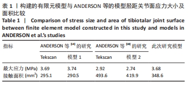
[14] ANDERSON DD, GOLDSWORTHY JK, LI W, et al. Physical validation of a patient-specific contact finite element model of the ankle. J Biomech. 2007;40(8): 1662-1669.
[15] CHEUNG JT, AN KN, ZHANG M. Consequences of partial and total plantar fascia release: a finite element study. Foot Ankle Int. 2006;27(2):125-132.
[16] ANDERSON DD, GOLDSWORTHY JK, LI W, et al. Physical validation of a patient-specific contact finite element model of the ankle. J Biomech. 2007;40(8):1662-1669.
[17] 白子兴,曹旭含,孙承颐,等.微创治疗拇外翻术后绷带外固定:有限元分析截骨端稳定性[J].中国组织工程研究,2020,24(18):2811-2816.
[18] ANDERSON DD, GOLDSWORTHY JK, SHIVANNA K, et al. Intra-ar ticular contact stress distributions at the ankle throughout stance phase-patient-specific finite element analysis as a metric of degenerationpropensity. Biomech Model Mechanobiol. 2006; 5(2-3): 82-89.
[19] 刘清华,余斌,金丹,等.正常人足踝部有限元模型的构建研究[J].中华创伤骨科杂志,2010,12(2):174-177.
[20] KRISTEN KH, BERGER K, BERGER C, et al. The first metatarsal bone under loading conditions: a finite element analysis. Foot Ankle Clin. 2005;10(1):1-14.
[21] 谢强,王智慧,刘文一,等.踝关节融合三维有限元模型的初步建立及验证[J].中国骨与关节损伤杂志,2018,33(6):645-646.
[22] 冯其金,赵玲娟,郑昆仑,等.有限元分析法在腰椎生物力学中的研究进展[J].中国中西医结合外科杂志,2018,24(2):255-258.
[23] KOSIK KB, TERADA M, MCCANN R, et al. Differences in temporal gait mechanics are associated with decreased perceived ankle joint health in individuals with chronic ankle instability. Gait Posture. 2019;70:403-407.
[24] ANDREOPOULOU G, MAHAD DJ, MERCER TH, et al. Test-retest reliability and minimal detectable change of ankle kinematics and spatiotemporal parameters in MS population. Gait Posture. 2019;74:218-222.
[25] WASHABAUGH EP, AUGENSTEIN TE, KRISHNAN C. Functional resistance training during walking: Mode of application differentially affects gait biomechanics and muscle activation patterns. Gait Posture. 2020;75:129-136.
[26] PUREVSUREN T, KIM K, BATBAATAR M, et al. Influence of ankle joint plantarflexion and dorsiflexion on lateral ankle sprain: A computational study. Proc Inst Mech Eng H. 2018;232(5):458-467.
[27] FRYZOWICZ A, MURAWA M, KABACIŃSKI J, et al.Reference values of spatiotemporal parameters, joint angles, ground reaction forces, and plantar pressure distribution during normal gait in young women. Acta Bioeng Biomech. 2018;20(1):49-57.
[28] LOHRER H. Lateral ankle ligament bracing. Oper Orthop Traumatol. 2019,31(3): 191-200.
[29] BOTTLANG M,FITZPATRICK DC,SHEERIN D,et al.Dynamic fixation of distal femur fractures using far cortical locking screws:a prospective observational study. J Orthop Trauma. 2014;28(4):181-188
[30] Filardi V. Finite element analysis of the foot: Stress and displacement shielding. J Orthop. 2018;15(4):974-979.
[31] 黄萍,钟慧敏,陈博,等.正常青年人三维步态:时空及运动学和运动力学参数分析[J].中国组织工程研究,2015,19(24):3882-3888.
[32] O’NEIL JT, GUYTON GP. Revision of Surgical Lateral Ankle Ligament Stabilization. Foot Ankle Clin. 2018;23(4):605-624.
[33] WAIZY H, HARRASSER N, FEHSKE K. Lateral ligament injuries. Unfallchirurg. 2018; 121(9):683-692.
[34] 徐志庆,刘云鹏,华国军,等.前踝撞击征发生机制的生物力学有限元分析[J].中国矫形外科杂志,2017,25(14):1303-1307. |