中国组织工程研究 ›› 2022, Vol. 26 ›› Issue (3): 371-375.doi: 10.12307/2022.061

• 数字化骨科 digital orthopedics • 上一篇    下一篇

3D打印可控式张力带的张力与皮肤缺损模型鼠皮肤再生

买合木提•亚库甫,孙琴琴,陈洪涛,刘  旭,伊力亚尔•阿不都斯木,阿布都萨拉木•阿布都克力木,刘建疆   

  1. 新疆医科大学第六附属医院运动医学科,新疆维吾尔自治区乌鲁木齐市   830002
  • 收稿日期:2021-02-06 修回日期:2021-02-08 接受日期:2021-04-15 出版日期:2022-01-28 发布日期:2021-10-27
  • 通讯作者: 刘建疆,主任,主任医师,新疆医科大学第六附属医院运动医学科,新疆维吾尔自治区乌鲁木齐市 830002
  • 作者简介:买合木提•亚库甫,男,1987年生,新疆维吾尔自治区乌鲁木齐市人,2020年吉林大学毕业,博士,主治医师,主要从事数字骨科、3D打印技术方面的研究。
  • 基金资助:
    新疆维吾尔自治区自然科学基金(2016D01C223),项目负责人:买合木提•亚库甫

Tension of 3D printed controllable tension band and skin regeneration of skin defect model rats

Maihemuti•Yakufu, Sun Qinqin, Chen Hongtao, Liu Xu, Yiliyaer•Abudusimu, Abudushalamu•Abudukelimu, Liu Jianjiang   

  1. Department of Sports Medicine, Sixth Affiliated Hospital of Xinjiang Medical University, Urumqi 830002, Xinjiang Uygur Autonomous Region, China
  • Received:2021-02-06 Revised:2021-02-08 Accepted:2021-04-15 Online:2022-01-28 Published:2021-10-27
  • Contact: Liu Jianjiang, Chief physician, Department of Sports Medicine, Sixth Affiliated Hospital of Xinjiang Medical University, Urumqi 830002, Xinjiang Uygur Autonomous Region, China
  • About author:Maihemuti•Yakufu, MD, Attending physician, Department of Sports Medicine, Sixth Affiliated Hospital of Xinjiang Medical University, Urumqi 830002, Xinjiang Uygur Autonomous Region, China
  • Supported by:
    the Natural Science Foundation of Xinjiang Uygur Autonomous Region, No. 2016D01C223 (to MY)

摘要:

文题释义:
皮肤缺损:为创伤、烧伤、肿瘤等原因导致皮肤组织的缺失,目前临床上常用的治疗方法包括皮肤移植、皮瓣移植等。
绑鞋带技术:是皮肤缺损创面的一种治疗方法,类似于鞋带一样,缝线或硅胶线从创面两侧来回相互交叉穿过,能够将缝线的张力均匀分散到皮肤缺损创面边缘,有效刺激皮肤边缘增殖,从而治疗皮肤缺损。

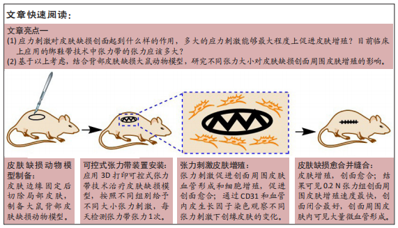
背景:虽然张力带技术能够刺激皮肤再生修复皮肤缺损,被成功应用于临床取得了满意的效果,但最适宜皮肤再生的张力大小尚无定论。
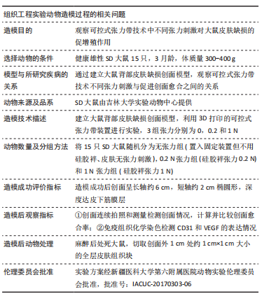
目的:探讨不同张力刺激对皮肤缺损模型大鼠皮肤再生的影响。
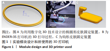
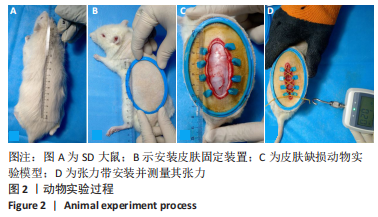
方法:选用雄性3月龄SD大鼠作为实验动物,通过3D打印的椭圆形皮肤固定装置固定SD大鼠背部皮肤,建立大鼠背部皮肤缺损创面模型,3D打印张力带装置固定在皮肤边缘,绑鞋带原理安装硅胶袢,并通过拉力检测器调整硅胶袢的不同张力参数(0,0.2,1 N)。造模术后当天、3 d和7 d观察测量皮肤缺损面积的变化;造模术后7 d取创面边缘皮肤组织学切片进行CD31和血管内皮生长因子免疫组织化学染色,观察不同张力刺激对皮肤再生的影响。
结果与结论:①适当大小的张力刺激能够有效刺激皮肤增殖愈合;无张力刺激的皮肤缺损创面会随着皮肤边缘的挛缩,创面进一步扩大,增加皮肤缺损面积,不利于皮肤缺损的治疗;②张力刺激不是越大越好,在同样时间点0.2 N张力组创面缩小速度比1 N张力组快,1 N张力组可见创面边缘皮肤硬化、弹性降低,考虑可能因为较大张力导致皮肤缺血所导致;③0.2 N张力组微血管数量明显增多,血管内皮生长因子表达量明显高于0 N张力组和1 N张力组,说明张力刺激能够促进微血管形成,能够提高血管内皮生长因子表达量,但是高张力刺激(此次研究中1 N)不利于皮肤缺损周围皮肤微血管形成,不利于创面愈合;④提示适当的张力刺激能够引起局部皮肤中广泛细胞外基质的重建;可通过诱导血管再生、改善局部细胞代谢所需营养物质的供应,促进细胞骨架重组,此过程对张力的大小有严格要求。
https://orcid.org/0000-0001-9853-1923 (买合木提•亚库甫) 

中国组织工程研究杂志出版内容重点:人工关节;骨植入物;脊柱;骨折;内固定;数字化骨科;组织工程

关键词: 3D打印, 可控式张力带, 皮肤缺损, 皮肤再生

Abstract: BACKGROUND: Although the tension band technique can stimulate skin regeneration to repair skin defects, it has been successfully applied in clinical practice and has achieved satisfactory results, but the most suitable tension intensity for skin regeneration is still inconclusive.  
OBJECTIVE: To discuss the effect of different tension intensities on skin regeneration in rat models of skin defect.
METHODS:  Male SD rats aged 3 months old were selected as experimental animals. The back skin of SD rats was fixed by the 3D printed oval skin fixing device to establish rat models of skin defect wound on the back, and the 3D printed tension band device was fixed on the edge of the skin. The silicone loop was installed on the principle of tying shoelaces, and different tension parameters (0, 0.2, 1 N) of the silicone loop were adjusted by the tension detector. The changes in the area of the skin defect were observed immediately, 3, and 7 days after model establishment. At 7 days after the surgical procedure, histological sections of the skin at the edge of the wound were taken for immunohistochemical staining of CD31 and vascular endothelial growth factor to observe the effect of different tension stimulation on skin regeneration.  
RESULTS AND CONCLUSION: (1) Appropriate tension stimulation could effectively stimulate skin proliferation and healing; skin defect wounds without tension stimulation could expand with the contracture of the skin edge and increase the area of the skin defect, which was not conducive to the treatment of skin defects. (2) Tension stimulation was not the bigger the better. Simultaneously, the wound shrinkage speed of 0.2 N tension group was faster than that of 1 N tension group. In 1 N tension group, the skin around the wound was sclerotic and the elasticity was decreased. It was considered that the larger tension led to skin ischemia. (3) The number of microvessels in the 0.2 N tension group was significantly increased, and the expression of vascular endothelial growth factor was significantly higher than that of the 0 N tension and 1 N tension groups, indicating that tension stimulation could promote the formation of microvessels and increase the expression of vascular endothelial growth factor, but high tension stimulation (1 N in this study) is not conducive to the formation of skin capillaries around the skin defect and is not conducive to wound healing. (4) It is concluded that appropriate tension can cause extensive reconstruction of extracellular matrix in local skin. By inducing angiogenesis, it can improve the supply of nutrients for local cell metabolism and promote the reorganization of cytoskeleton. This process has strict requirements on the size of tension.

Key words: 3D printing, controllable tension band, skin defect, skin regeneration

中图分类号: